 Movie
Movie Controller

[English] 日本語
 Yorodumi
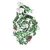
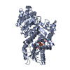
- PDB-7p0n: Crystal structure of L-Trp/Indoleamine 2,3-dioxygenagse 1 (hIDO1)... -
+ Open data

- Basic information
| Entry | Database: PDB / ID: 7p0n | ||||||
|---|---|---|---|---|---|---|---|
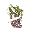
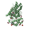
| Title | Crystal structure of L-Trp/Indoleamine 2,3-dioxygenagse 1 (hIDO1) complex with the JK-loop refined in the open conformation | ||||||
 Components Components | Indoleamine 2,3-dioxygenase 1 | ||||||
 Keywords Keywords | OXIDOREDUCTASE / L-Trp metabolism / hemoprotein / dynamics loop | ||||||
| Function / homology |  Function and homology information indoleamine 2,3-dioxygenase / smooth muscle contractile fiber / indoleamine 2,3-dioxygenase activity / positive regulation of chronic inflammatory response / kynurenic acid biosynthetic process / tryptophan 2,3-dioxygenase activity / positive regulation of T cell tolerance induction / tryptophan catabolic process to kynurenine / stereocilium bundle / positive regulation of type 2 immune response ... indoleamine 2,3-dioxygenase / smooth muscle contractile fiber / indoleamine 2,3-dioxygenase activity / positive regulation of chronic inflammatory response / kynurenic acid biosynthetic process / tryptophan 2,3-dioxygenase activity / positive regulation of T cell tolerance induction / tryptophan catabolic process to kynurenine / stereocilium bundle / positive regulation of type 2 immune response / 'de novo' NAD biosynthetic process from tryptophan / tryptophan catabolic process / Tryptophan catabolism / positive regulation of T cell apoptotic process / negative regulation of T cell apoptotic process / swimming behavior / negative regulation of interleukin-10 production / multicellular organismal response to stress / T cell proliferation / negative regulation of T cell proliferation / positive regulation of interleukin-12 production / female pregnancy / response to lipopolysaccharide / electron transfer activity / inflammatory response / heme binding / metal ion binding / cytosol / cytoplasm Function and homology information indoleamine 2,3-dioxygenase / smooth muscle contractile fiber / indoleamine 2,3-dioxygenase activity / positive regulation of chronic inflammatory response / kynurenic acid biosynthetic process / tryptophan 2,3-dioxygenase activity / positive regulation of T cell tolerance induction / tryptophan catabolic process to kynurenine / stereocilium bundle / positive regulation of type 2 immune response ... indoleamine 2,3-dioxygenase / smooth muscle contractile fiber / indoleamine 2,3-dioxygenase activity / positive regulation of chronic inflammatory response / kynurenic acid biosynthetic process / tryptophan 2,3-dioxygenase activity / positive regulation of T cell tolerance induction / tryptophan catabolic process to kynurenine / stereocilium bundle / positive regulation of type 2 immune response / 'de novo' NAD biosynthetic process from tryptophan / tryptophan catabolic process / Tryptophan catabolism / positive regulation of T cell apoptotic process / negative regulation of T cell apoptotic process / swimming behavior / negative regulation of interleukin-10 production / multicellular organismal response to stress / T cell proliferation / negative regulation of T cell proliferation / positive regulation of interleukin-12 production / female pregnancy / response to lipopolysaccharide / electron transfer activity / inflammatory response / heme binding / metal ion binding / cytosol / cytoplasmSimilarity search - Function | ||||||
| Biological species |  Homo sapiens (human) Homo sapiens (human) | ||||||
| Method | X-RAY DIFFRACTION / SYNCHROTRON / MOLECULAR REPLACEMENT / Resolution: 2.5 Å | ||||||
 Authors Authors | Mirgaux, M. / Wouters, J. | ||||||
 Citation Citation | Journal: Int J Tryptophan Res / Year: 2021 Title: Temporary Intermediates of L-Trp Along the Reaction Pathway of Human Indoleamine 2,3-Dioxygenase 1 and Identification of an Exo Site. Authors: Mirgaux, M. / Leherte, L. / Wouters, J. | ||||||
| History |
|
- Structure visualization
| Structure viewer | Molecule: MolmilJmol/JSmol |
|---|
- Downloads & links
-Download
| PDBx/mmCIF format | 7p0n.cif.gz | 396.5 KB | Display | PDBx/mmCIF format |
|---|---|---|---|---|
| PDB format | pdb7p0n.ent.gz | 262.7 KB | Display | PDB format |
| PDBx/mmJSON format | 7p0n.json.gz | Tree view | PDBx/mmJSON format | |
| Others |  Other downloads Other downloads |
-Validation report
| Arichive directory | https://data.pdbj.org/pub/pdb/validation_reports/p0/7p0nftp://data.pdbj.org/pub/pdb/validation_reports/p0/7p0n | HTTPS FTP |
|---|
-Related structure data
| Related structure data |  7ngeC 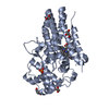 7p0rC  7a62S S: Starting model for refinement C: citing same article ( |
|---|---|
| Similar structure data |


-Links
 PDBj
PDBj


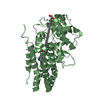
- Assembly
| Deposited unit | 
| ||||||||||||
|---|---|---|---|---|---|---|---|---|---|---|---|---|---|
| 1 | 
| ||||||||||||
| 2 | 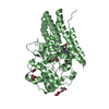
| ||||||||||||
| 3 | 
| ||||||||||||
| 4 | 
| ||||||||||||
| Unit cell |
| ||||||||||||
| Components on special symmetry positions |
|
-Components
-Protein , 1 types, 4 molecules ABCD
| #1: Protein | / IDO-1 / Indoleamine-pyrrole 2 / 3-dioxygenase Mass: 45300.898 Da / Num. of mol.: 4 / Mutation: K116A, K117A Source method: isolated from a genetically manipulated source Source: (gene. exp.) Homo sapiens (human) / Gene: IDO1, IDO, INDO / Plasmid: pet28-a / Details (production host): Mutations K116A, K117A / Production host:  Escherichia coli BL21(DE3) (bacteria) / References: UniProt: P14902, indoleamine 2,3-dioxygenase Escherichia coli BL21(DE3) (bacteria) / References: UniProt: P14902, indoleamine 2,3-dioxygenase |
|---|
-Non-polymers , 8 types, 531 molecules 
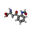


| #2: Chemical | ChemComp-HEM / Heme B Mass: 616.487 Da / Num. of mol.: 4 / Source method: obtained synthetically / Formula: C34H32FeN4O4 / Feature type: SUBJECT OF INVESTIGATION Mass: 616.487 Da / Num. of mol.: 4 / Source method: obtained synthetically / Formula: C34H32FeN4O4 / Feature type: SUBJECT OF INVESTIGATION#3: Chemical | ChemComp-NFK / N'-Formylkynurenine Mass: 236.224 Da / Num. of mol.: 4 / Source method: obtained synthetically / Formula: C11H12N2O4 / Feature type: SUBJECT OF INVESTIGATION Mass: 236.224 Da / Num. of mol.: 4 / Source method: obtained synthetically / Formula: C11H12N2O4 / Feature type: SUBJECT OF INVESTIGATION#4: Chemical | Phosphate Mass: 94.971 Da / Num. of mol.: 2 / Source method: obtained synthetically / Formula: PO4 Mass: 94.971 Da / Num. of mol.: 2 / Source method: obtained synthetically / Formula: PO4#5: Chemical | ChemComp-GOL / Glycerol Mass: 92.094 Da / Num. of mol.: 21 / Source method: obtained synthetically / Formula: C3H8O3 Mass: 92.094 Da / Num. of mol.: 21 / Source method: obtained synthetically / Formula: C3H8O3#6: Chemical | Oxygen Mass: 31.999 Da / Num. of mol.: 3 / Source method: obtained synthetically / Formula: O2 Mass: 31.999 Da / Num. of mol.: 3 / Source method: obtained synthetically / Formula: O2#7: Chemical | Tryptophan Type: L-peptide linking / Mass: 204.225 Da / Num. of mol.: 3 / Source method: obtained synthetically / Formula: C11H12N2O2 / Feature type: SUBJECT OF INVESTIGATION Type: L-peptide linking / Mass: 204.225 Da / Num. of mol.: 3 / Source method: obtained synthetically / Formula: C11H12N2O2 / Feature type: SUBJECT OF INVESTIGATION#8: Chemical | Chloride Mass: 35.453 Da / Num. of mol.: 2 / Source method: obtained synthetically / Formula: Cl Mass: 35.453 Da / Num. of mol.: 2 / Source method: obtained synthetically / Formula: Cl#9: Water | ChemComp-HOH / | WaterMass: 18.015 Da / Num. of mol.: 492 / Source method: isolated from a natural source / Formula: H2O |
|---|
-Details
| Has ligand of interest | Y |
|---|
-Experimental details
-Experiment
| Experiment | Method: X-RAY DIFFRACTION / Number of used crystals: 1 |
|---|
- Sample preparation
| Crystal | Density Matthews: 2.81 Å3/Da / Density % sol: 56.72 % / Description: Red rectangular crystal 0.3mm |
|---|---|
| Crystal grow | Temperature: 298 K / Method: vapor diffusion, hanging drop / pH: 6.4 Details: 14.5% PEG 3350 0.1M Phosphate buffer 2mM L-Trp Protein buffer : Hepes 5mM, 200mM NaCl, 5mM DTT Cryoprotection: 20 mM sodium dithionite, 20% glycerol PH range: 6.0-6.4 / Temp details: Room temperature |
-Data collection
| Diffraction | Mean temperature: 100 K / Serial crystal experiment: N |
|---|---|
| Diffraction source | Source: SYNCHROTRON / Site: SOLEIL  / Beamline: PROXIMA 2 / Wavelength: 0.98012 Å / Beamline: PROXIMA 2 / Wavelength: 0.98012 Å |
| Detector | Type: DECTRIS EIGER2 X 9M / Detector: PIXEL / Date: Jul 5, 2020 |
| Radiation | Monochromator: SI(111) / Protocol: SINGLE WAVELENGTH / Monochromatic (M) / Laue (L): M / Scattering type: x-ray |
| Radiation wavelength | Wavelength: 0.98012 Å / Relative weight: 1 |
| Reflection | Resolution: 2.5→49.29 Å / Num. obs: 71786 / % possible obs: 99.34 % / Redundancy: 7 % / Biso Wilson estimate: 52.26 Å2 / CC1/2: 0.995 / CC star: 0.999 / Net I/σ(I): 6.7 |
| Reflection shell | Resolution: 2.504→2.593 Å / Mean I/σ(I) obs: 0.95 / Num. unique obs: 7022 / CC1/2: 0.606 / CC star: 0.869 / % possible all: 97.88 |
- Processing
| Software |
| |||||||||||||||||||||||||||||||||||||||||||||||||||||||||||||||||||||||||||||||||||||||||||||||||||||||||||||||||||||||||||||||||||||||||||||||||||||||||||||||||||||||||||||||||||||||||||||
|---|---|---|---|---|---|---|---|---|---|---|---|---|---|---|---|---|---|---|---|---|---|---|---|---|---|---|---|---|---|---|---|---|---|---|---|---|---|---|---|---|---|---|---|---|---|---|---|---|---|---|---|---|---|---|---|---|---|---|---|---|---|---|---|---|---|---|---|---|---|---|---|---|---|---|---|---|---|---|---|---|---|---|---|---|---|---|---|---|---|---|---|---|---|---|---|---|---|---|---|---|---|---|---|---|---|---|---|---|---|---|---|---|---|---|---|---|---|---|---|---|---|---|---|---|---|---|---|---|---|---|---|---|---|---|---|---|---|---|---|---|---|---|---|---|---|---|---|---|---|---|---|---|---|---|---|---|---|---|---|---|---|---|---|---|---|---|---|---|---|---|---|---|---|---|---|---|---|---|---|---|---|---|---|---|---|---|---|---|---|---|
| Refinement | Method to determine structure: MOLECULAR REPLACEMENT Starting model: 7A62 Resolution: 2.5→49.29 Å / SU ML: 0.3948 / Cross valid method: FREE R-VALUE / σ(F): 1.34 / Phase error: 35.0264 Stereochemistry target values: GeoStd + Monomer Library + CDL v1.2
| |||||||||||||||||||||||||||||||||||||||||||||||||||||||||||||||||||||||||||||||||||||||||||||||||||||||||||||||||||||||||||||||||||||||||||||||||||||||||||||||||||||||||||||||||||||||||||||
| Solvent computation | Shrinkage radii: 0.9 Å / VDW probe radii: 1.11 Å / Solvent model: FLAT BULK SOLVENT MODEL | |||||||||||||||||||||||||||||||||||||||||||||||||||||||||||||||||||||||||||||||||||||||||||||||||||||||||||||||||||||||||||||||||||||||||||||||||||||||||||||||||||||||||||||||||||||||||||||
| Displacement parameters | Biso mean: 58.73 Å2 | |||||||||||||||||||||||||||||||||||||||||||||||||||||||||||||||||||||||||||||||||||||||||||||||||||||||||||||||||||||||||||||||||||||||||||||||||||||||||||||||||||||||||||||||||||||||||||||
| Refinement step | Cycle: LAST / Resolution: 2.5→49.29 Å
| |||||||||||||||||||||||||||||||||||||||||||||||||||||||||||||||||||||||||||||||||||||||||||||||||||||||||||||||||||||||||||||||||||||||||||||||||||||||||||||||||||||||||||||||||||||||||||||
| Refine LS restraints |
| |||||||||||||||||||||||||||||||||||||||||||||||||||||||||||||||||||||||||||||||||||||||||||||||||||||||||||||||||||||||||||||||||||||||||||||||||||||||||||||||||||||||||||||||||||||||||||||
| LS refinement shell |
|
