 Movie
Movie Controller

[English] 日本語
 Yorodumi
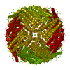
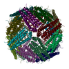
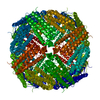
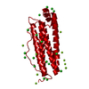
- PDB-7o67: Crystal structure of human mitochondrial ferritin (hMTF) Fe(II)-l... -
+ Open data

- Basic information
| Entry | Database: PDB / ID: 7o67 | ||||||
|---|---|---|---|---|---|---|---|
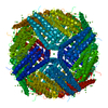
| Title | Crystal structure of human mitochondrial ferritin (hMTF) Fe(II)-loaded for 15 minutes showing either a dioxygen or a superoxide anion coordinated to iron ions in the ferroxidase site | ||||||
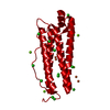
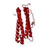
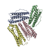
 Components Components | Ferritin, mitochondrial | ||||||
 Keywords Keywords | OXIDOREDUCTASE / human mitochondrial ferritin / hMTF / time-controlled iron loading / ferroxidase site / dioxygen / superoxide anion | ||||||
| Function / homology |  Function and homology information Function and homology informationpositive regulation of lyase activity / positive regulation of succinate dehydrogenase activity / positive regulation of aconitate hydratase activity / ferroxidase / intracellular sequestering of iron ion / ferroxidase activity / ferric iron binding / ferrous iron binding / Iron uptake and transport / iron ion transport ...positive regulation of lyase activity / positive regulation of succinate dehydrogenase activity / positive regulation of aconitate hydratase activity / ferroxidase / intracellular sequestering of iron ion / ferroxidase activity / ferric iron binding / ferrous iron binding / Iron uptake and transport / iron ion transport / intracellular iron ion homeostasis / mitochondrial matrix / iron ion binding / positive regulation of cell population proliferation / mitochondrion / nucleus / cytoplasmSimilarity search - Function | ||||||
| Biological species |  Homo sapiens (human) Homo sapiens (human) | ||||||
| Method | X-RAY DIFFRACTION / SYNCHROTRON / MOLECULAR REPLACEMENT / Resolution: 1.86 Å | ||||||
 Authors Authors | Pozzi, C. / Ciambellotti, S. / Tassone, G. / Turano, P. / Mangani, S. | ||||||
 Citation Citation | Journal: Chemistry / Year: 2021 Title: Iron Binding in the Ferroxidase Site of Human Mitochondrial Ferritin. Authors: Ciambellotti, S. / Pratesi, A. / Tassone, G. / Turano, P. / Mangani, S. / Pozzi, C. | ||||||
| History |
|
- Structure visualization
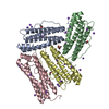
| Structure viewer | Molecule: MolmilJmol/JSmol |
|---|
- Downloads & links
-Download
| PDBx/mmCIF format | 7o67.cif.gz | 59.7 KB | Display | PDBx/mmCIF format |
|---|---|---|---|---|
| PDB format | pdb7o67.ent.gz | 43 KB | Display | PDB format |
| PDBx/mmJSON format | 7o67.json.gz | Tree view | PDBx/mmJSON format | |
| Others |  Other downloads Other downloads |
-Validation report
| Arichive directory | https://data.pdbj.org/pub/pdb/validation_reports/o6/7o67ftp://data.pdbj.org/pub/pdb/validation_reports/o6/7o67 | HTTPS FTP |
|---|
-Related structure data
| Related structure data |  7o63C  7o64C  7o65C  7o66C  7o68C  7o69C  7o6aC 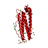 7o6cC  7o6dC  7owyC  1r03S C: citing same article ( S: Starting model for refinement |
|---|---|
| Similar structure data |


-Links
 PDBj
PDBj


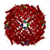
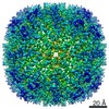
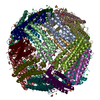
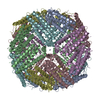
- Assembly
| Deposited unit | 
| ||||||||||||||||||||||||||||||
|---|---|---|---|---|---|---|---|---|---|---|---|---|---|---|---|---|---|---|---|---|---|---|---|---|---|---|---|---|---|---|---|
| 1 | x 24
| ||||||||||||||||||||||||||||||
| Unit cell |
| ||||||||||||||||||||||||||||||
| Components on special symmetry positions |
|
-Components
| #1: Protein | Mass: 21107.568 Da / Num. of mol.: 1 Source method: isolated from a genetically manipulated source Source: (gene. exp.) Homo sapiens (human) / Gene: FTMT / Plasmid: pET-3a / Production host:  Escherichia coli BL21(DE3) (bacteria) / Variant (production host): -pLysS / References: UniProt: Q8N4E7, ferroxidase Escherichia coli BL21(DE3) (bacteria) / Variant (production host): -pLysS / References: UniProt: Q8N4E7, ferroxidase | ||||||
|---|---|---|---|---|---|---|---|
| #2: Chemical | ChemComp-OXY / Oxygen  Mass: 31.999 Da / Num. of mol.: 1 / Source method: obtained synthetically / Formula: O2 Mass: 31.999 Da / Num. of mol.: 1 / Source method: obtained synthetically / Formula: O2 | ||||||
| #3: Chemical | ChemComp-FE2 /   Mass: 55.845 Da / Num. of mol.: 9 / Source method: obtained synthetically / Formula: Fe / Feature type: SUBJECT OF INVESTIGATION Mass: 55.845 Da / Num. of mol.: 9 / Source method: obtained synthetically / Formula: Fe / Feature type: SUBJECT OF INVESTIGATION#4: Chemical | ChemComp-CL / | Chloride  Mass: 35.453 Da / Num. of mol.: 1 / Source method: obtained synthetically / Formula: Cl Mass: 35.453 Da / Num. of mol.: 1 / Source method: obtained synthetically / Formula: Cl#5: Water | ChemComp-HOH / | Water Mass: 18.015 Da / Num. of mol.: 233 / Source method: isolated from a natural source / Formula: H2O Mass: 18.015 Da / Num. of mol.: 233 / Source method: isolated from a natural source / Formula: H2OHas ligand of interest | Y | |
-Experimental details
-Experiment
| Experiment | Method: X-RAY DIFFRACTION / Number of used crystals: 1 |
|---|
- Sample preparation
| Crystal | Density Matthews: 3.16 Å3/Da / Density % sol: 61.12 % / Description: Octahedral crystals |
|---|---|
| Crystal grow | Temperature: 281.15 K / Method: vapor diffusion, hanging drop / Details: 1.6-2 M MgCl2 6H2O and 0.1 M bicine pH 9.0 |
-Data collection
| Diffraction |
| ||||||||||||||||||||||||||||||||||||||||||||
|---|---|---|---|---|---|---|---|---|---|---|---|---|---|---|---|---|---|---|---|---|---|---|---|---|---|---|---|---|---|---|---|---|---|---|---|---|---|---|---|---|---|---|---|---|---|
| Diffraction source |
| ||||||||||||||||||||||||||||||||||||||||||||
| Detector |
| ||||||||||||||||||||||||||||||||||||||||||||
| Radiation |
| ||||||||||||||||||||||||||||||||||||||||||||
| Radiation wavelength |
| ||||||||||||||||||||||||||||||||||||||||||||
| Reflection | Entry-ID: 7O67 / Observed criterion σ(F): 2
| ||||||||||||||||||||||||||||||||||||||||||||
| Reflection shell |
|

- Processing
| Software |
| |||||||||||||||||||||||||||||||||||||||||||||
|---|---|---|---|---|---|---|---|---|---|---|---|---|---|---|---|---|---|---|---|---|---|---|---|---|---|---|---|---|---|---|---|---|---|---|---|---|---|---|---|---|---|---|---|---|---|---|
| Refinement | Method to determine structure: MOLECULAR REPLACEMENT Starting model: 1R03 Resolution: 1.86→46.09 Å / Cor.coef. Fo:Fc: 0.964 / Cor.coef. Fo:Fc free: 0.944 / SU B: 2.84 / SU ML: 0.082 / Cross valid method: THROUGHOUT / σ(F): 0 / ESU R: 0.11 / ESU R Free: 0.119 / Stereochemistry target values: MAXIMUM LIKELIHOOD / Details: U VALUES : REFINED INDIVIDUALLY
| |||||||||||||||||||||||||||||||||||||||||||||
| Solvent computation | Ion probe radii: 0.8 Å / Shrinkage radii: 0.8 Å / VDW probe radii: 1.2 Å / Solvent model: MASK | |||||||||||||||||||||||||||||||||||||||||||||
| Displacement parameters | Biso max: 85.28 Å2 / Biso mean: 27.859 Å2 / Biso min: 16.21 Å2
| |||||||||||||||||||||||||||||||||||||||||||||
| Refine analyze | Luzzati coordinate error obs: 0.2044 Å | |||||||||||||||||||||||||||||||||||||||||||||
| Refinement step | Cycle: final / Resolution: 1.86→46.09 Å
| |||||||||||||||||||||||||||||||||||||||||||||
| Refine LS restraints |
| |||||||||||||||||||||||||||||||||||||||||||||
| LS refinement shell | Resolution: 1.86→1.908 Å / Rfactor Rfree error: 0 / Total num. of bins used: 20
|
