 Movie
Movie Controller

[English] 日本語
 Yorodumi
- PDB-5n26: X-ray structure of human heavy chain ferritin in complex with cis... -
+ Open data

- Basic information
| Entry | Database: PDB / ID: 5n26 | ||||||
|---|---|---|---|---|---|---|---|
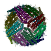
| Title | X-ray structure of human heavy chain ferritin in complex with cisplatin | ||||||
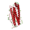
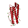
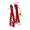
 Components Components | Ferritin heavy chain | ||||||
 Keywords Keywords | OXIDOREDUCTASE / ferritin | ||||||
| Function / homology |  Function and homology information Function and homology informationiron ion sequestering activity / : / autolysosome / Scavenging by Class A Receptors / Golgi Associated Vesicle Biogenesis / ferroxidase / intracellular sequestering of iron ion / ferroxidase activity / negative regulation of fibroblast proliferation / ferric iron binding ...iron ion sequestering activity / : / autolysosome / Scavenging by Class A Receptors / Golgi Associated Vesicle Biogenesis / ferroxidase / intracellular sequestering of iron ion / ferroxidase activity / negative regulation of fibroblast proliferation / ferric iron binding / ferrous iron binding / Iron uptake and transport / tertiary granule lumen / iron ion transport / intracellular iron ion homeostasis / ficolin-1-rich granule lumen / immune response / iron ion binding / negative regulation of cell population proliferation / Neutrophil degranulation / extracellular exosome / extracellular region / identical protein binding / nucleus / cytosol / cytoplasmSimilarity search - Function | ||||||
| Biological species |  Homo sapiens (human) Homo sapiens (human) | ||||||
| Method | X-RAY DIFFRACTION / MOLECULAR REPLACEMENT / Resolution: 2.05 Å | ||||||
 Authors Authors | Ferraro, G. / Merlino, A. | ||||||
 Citation Citation | Journal: Inorg Chem / Year: 2017 Title: Cisplatin Binding Sites in Human H-Chain Ferritin. Authors: Ferraro, G. / Ciambellotti, S. / Messori, L. / Merlino, A. | ||||||
| History |
|
- Structure visualization
| Structure viewer | Molecule: MolmilJmol/JSmol |
|---|
- Downloads & links
-Download
| PDBx/mmCIF format | 5n26.cif.gz | 60.2 KB | Display | PDBx/mmCIF format |
|---|---|---|---|---|
| PDB format | pdb5n26.ent.gz | 43.7 KB | Display | PDB format |
| PDBx/mmJSON format | 5n26.json.gz | Tree view | PDBx/mmJSON format | |
| Others |  Other downloads Other downloads |
-Validation report
| Arichive directory | https://data.pdbj.org/pub/pdb/validation_reports/n2/5n26ftp://data.pdbj.org/pub/pdb/validation_reports/n2/5n26 | HTTPS FTP |
|---|
-Related structure data
| Related structure data |  5n27C 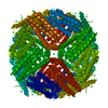 4y08S S: Starting model for refinement C: citing same article ( |
|---|---|
| Similar structure data |


-Links
 PDBj
PDBj


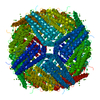
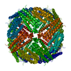
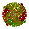
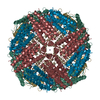
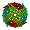
- Assembly
| Deposited unit | 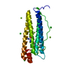
| ||||||||||||||||||||||||
|---|---|---|---|---|---|---|---|---|---|---|---|---|---|---|---|---|---|---|---|---|---|---|---|---|---|
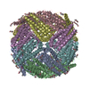
| 1 | x 24
| ||||||||||||||||||||||||
| Unit cell |
| ||||||||||||||||||||||||
| Components on special symmetry positions |
|
-Components
-Protein , 1 types, 1 molecules A
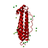
| #1: Protein | / Ferritin H subunit / Cell proliferation-inducing gene 15 protein Mass: 21124.459 Da / Num. of mol.: 1 Source method: isolated from a genetically manipulated source Source: (gene. exp.) Homo sapiens (human) / Gene: FTH1, FTH, FTHL6, OK/SW-cl.84, PIG15 / Production host:  Escherichia coli BL21(DE3) (bacteria) / References: UniProt: P02794, ferroxidase Escherichia coli BL21(DE3) (bacteria) / References: UniProt: P02794, ferroxidase |
|---|
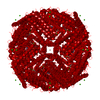
-Non-polymers , 5 types, 238 molecules 


| #2: Chemical | Cisplatin Mass: 300.045 Da / Num. of mol.: 3 / Source method: obtained synthetically / Formula: Cl2H6N2Pt / Comment: medication, chemotherapy*YM Mass: 300.045 Da / Num. of mol.: 3 / Source method: obtained synthetically / Formula: Cl2H6N2Pt / Comment: medication, chemotherapy*YM#3: Chemical | ChemComp-73M / |  Mass: 279.584 Da / Num. of mol.: 1 / Source method: obtained synthetically / Formula: ClH5N2OPt Mass: 279.584 Da / Num. of mol.: 1 / Source method: obtained synthetically / Formula: ClH5N2OPt#4: Chemical | ChemComp-CL / Chloride Mass: 35.453 Da / Num. of mol.: 6 / Source method: obtained synthetically / Formula: Cl Mass: 35.453 Da / Num. of mol.: 6 / Source method: obtained synthetically / Formula: Cl#5: Chemical | ChemComp-MG /  Mass: 24.305 Da / Num. of mol.: 5 / Source method: obtained synthetically / Formula: Mg Mass: 24.305 Da / Num. of mol.: 5 / Source method: obtained synthetically / Formula: Mg#6: Water | ChemComp-HOH / | WaterMass: 18.015 Da / Num. of mol.: 223 / Source method: isolated from a natural source / Formula: H2O |
|---|
-Experimental details
-Experiment
| Experiment | Method: X-RAY DIFFRACTION / Number of used crystals: 1 |
|---|
- Sample preparation
| Crystal | Density Matthews: 3.03 Å3/Da / Density % sol: 59.34 % |
|---|---|
| Crystal grow | Temperature: 293 K / Method: vapor diffusion, hanging drop / pH: 9 / Details: 2.0 M Magnesium chloride, 0.1 M bicine pH 9.0 |
-Data collection
| Diffraction | Mean temperature: 100 K |
|---|---|
| Diffraction source | Source: ROTATING ANODE / Type: RIGAKU MICROMAX-007 HF / Wavelength: 1.5418 Å |
| Detector | Type: RIGAKU SATURN 944 / Detector: CCD / Date: Sep 15, 2006 / Details: mirrors |
| Radiation | Protocol: SINGLE WAVELENGTH / Monochromatic (M) / Laue (L): M / Scattering type: x-ray |
| Radiation wavelength | Wavelength: 1.5418 Å / Relative weight: 1 |
| Reflection | Resolution: 2.05→105.7 Å / Num. obs: 16819 / % possible obs: 98.5 % / Observed criterion σ(F): 0 / Observed criterion σ(I): 0 / Redundancy: 7.9 % / Rmerge(I) obs: 0.143 / Rpim(I) all: 0.053 / Net I/σ(I): 17.8 |
| Reflection shell | Resolution: 2.05→2.09 Å / Redundancy: 6.9 % / Rmerge(I) obs: 0.588 / Mean I/σ(I) obs: 3.7 / CC1/2: 0.837 / Rpim(I) all: 0.236 / % possible all: 91.8 |
- Processing
| Software |
| ||||||||||||||||||||||||||||||||||||||||||||||||||||||||||||||||||||||||||||||||||||||||||||||||||||||||||||||||||||||||||||||||||||||||||||||||||||||||||||||||||||||||||||||||||||||
|---|---|---|---|---|---|---|---|---|---|---|---|---|---|---|---|---|---|---|---|---|---|---|---|---|---|---|---|---|---|---|---|---|---|---|---|---|---|---|---|---|---|---|---|---|---|---|---|---|---|---|---|---|---|---|---|---|---|---|---|---|---|---|---|---|---|---|---|---|---|---|---|---|---|---|---|---|---|---|---|---|---|---|---|---|---|---|---|---|---|---|---|---|---|---|---|---|---|---|---|---|---|---|---|---|---|---|---|---|---|---|---|---|---|---|---|---|---|---|---|---|---|---|---|---|---|---|---|---|---|---|---|---|---|---|---|---|---|---|---|---|---|---|---|---|---|---|---|---|---|---|---|---|---|---|---|---|---|---|---|---|---|---|---|---|---|---|---|---|---|---|---|---|---|---|---|---|---|---|---|---|---|---|---|
| Refinement | Method to determine structure: MOLECULAR REPLACEMENT Starting model: 4Y08 Resolution: 2.05→105.69 Å / Cor.coef. Fo:Fc: 0.956 / Cor.coef. Fo:Fc free: 0.937 / SU B: 3.406 / SU ML: 0.092 / Cross valid method: THROUGHOUT / ESU R: 0.149 / ESU R Free: 0.142 / Details: HYDROGENS HAVE BEEN ADDED IN THE RIDING POSITIONS
| ||||||||||||||||||||||||||||||||||||||||||||||||||||||||||||||||||||||||||||||||||||||||||||||||||||||||||||||||||||||||||||||||||||||||||||||||||||||||||||||||||||||||||||||||||||||
| Solvent computation | Ion probe radii: 0.8 Å / Shrinkage radii: 0.8 Å / VDW probe radii: 1.2 Å | ||||||||||||||||||||||||||||||||||||||||||||||||||||||||||||||||||||||||||||||||||||||||||||||||||||||||||||||||||||||||||||||||||||||||||||||||||||||||||||||||||||||||||||||||||||||
| Displacement parameters | Biso mean: 19.358 Å2
| ||||||||||||||||||||||||||||||||||||||||||||||||||||||||||||||||||||||||||||||||||||||||||||||||||||||||||||||||||||||||||||||||||||||||||||||||||||||||||||||||||||||||||||||||||||||
| Refinement step | Cycle: 1 / Resolution: 2.05→105.69 Å
| ||||||||||||||||||||||||||||||||||||||||||||||||||||||||||||||||||||||||||||||||||||||||||||||||||||||||||||||||||||||||||||||||||||||||||||||||||||||||||||||||||||||||||||||||||||||
| Refine LS restraints |
|