 Movie
Movie Controller


+ Open data

- Basic information
| Entry | Database: PDB / ID: 1r03 | ||||||
|---|---|---|---|---|---|---|---|
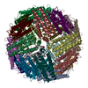
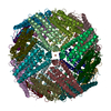
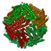
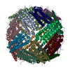
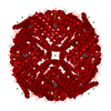
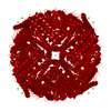
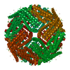
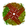
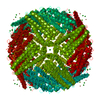
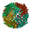
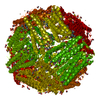
| Title | crystal structure of a human mitochondrial ferritin | ||||||
 Components Components | mitochondrial ferritin | ||||||
 Keywords Keywords | METAL BINDING PROTEIN / iron storage / ferritin / x-ray crystallography | ||||||
| Function / homology |  Function and homology information Function and homology informationpositive regulation of lyase activity / positive regulation of succinate dehydrogenase activity / positive regulation of aconitate hydratase activity / ferroxidase / intracellular sequestering of iron ion / ferroxidase activity / ferric iron binding / Iron uptake and transport / ferrous iron binding / iron ion transport ...positive regulation of lyase activity / positive regulation of succinate dehydrogenase activity / positive regulation of aconitate hydratase activity / ferroxidase / intracellular sequestering of iron ion / ferroxidase activity / ferric iron binding / Iron uptake and transport / ferrous iron binding / iron ion transport / intracellular iron ion homeostasis / mitochondrial matrix / iron ion binding / positive regulation of cell population proliferation / mitochondrion / nucleus / cytoplasmSimilarity search - Function | ||||||
| Biological species |  Homo sapiens (human) Homo sapiens (human) | ||||||
| Method | X-RAY DIFFRACTION / SYNCHROTRON / MOLECULAR REPLACEMENT / Resolution: 1.7 Å | ||||||
 Authors Authors | Corsi, B. / Santambrogio, P. / Arosio, P. / Levi, S. / Langlois d'Estaintot, B. / Granier, T. / Gallois, B. / Chevallier, J.M. / Precigoux, G. | ||||||
 Citation Citation | Journal: J.Mol.Biol. / Year: 2004 Title: Crystal Structure and Biochemical Properties of the Human Mitochondrial Ferritin and its Mutant Ser144Ala Authors: Langlois d'Estaintot, B. / Santambrogio, P. / Granier, T. / Gallois, B. / Chevallier, J.M. / Precigoux, G. / Levi, S. / Arosio, P. #1: Journal: J.Biol.Chem. / Year: 2001Title: A human mitochondrial ferritin encoded by an intronless gene Authors: Levi, S. / Corsi, B. / Bosisio, M. / Invernizzi, R. / Volz, A. / Sanford, D. / Arosio, P. / Drysdale, J. #2: Journal: J.Mol.Biol. / Year: 1997Title: Comparison of the three-dimensional structures of recombinant human H and horse L ferritins at high resolution ferritins at high resolution Authors: Hempstead, P.D. / Yewdall, S.J. / Alistair, R. / Lawson, D.M. / Artymiuk, P.J. / Rice, D.W. / Ford, G.C. / Harrison, P.M. | ||||||
| History |
|
- Structure visualization
| Structure viewer | Molecule: MolmilJmol/JSmol |
|---|
- Downloads & links
-Download
| PDBx/mmCIF format | 1r03.cif.gz | 59.7 KB | Display | PDBx/mmCIF format |
|---|---|---|---|---|
| PDB format | pdb1r03.ent.gz | 42.3 KB | Display | PDB format |
| PDBx/mmJSON format | 1r03.json.gz | Tree view | PDBx/mmJSON format | |
| Others |  Other downloads Other downloads |
-Validation report
| Arichive directory | https://data.pdbj.org/pub/pdb/validation_reports/r0/1r03ftp://data.pdbj.org/pub/pdb/validation_reports/r0/1r03 | HTTPS FTP |
|---|
-Related structure data
| Related structure data |  2fhaS S: Starting model for refinement |
|---|---|
| Similar structure data |


-Links
 PDBj
PDBj


- Assembly
| Deposited unit | 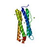
| ||||||||||||||||||
|---|---|---|---|---|---|---|---|---|---|---|---|---|---|---|---|---|---|---|---|
| 1 | x 24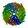
| ||||||||||||||||||
| Unit cell |
| ||||||||||||||||||
| Components on special symmetry positions |
| ||||||||||||||||||
| Details | coordinates for a complete multimer representing the known biologically significant oligomerization state of the molecule can be generated by applying the the symmetry operations: -x,-y,z; -x,y,-z; x,-y,-z; z,x,y; z,-x,-y; -z,-x,y; -z,x,-y; y,z,x; -y,z,-x; y,-z,-x; -y,-z,x; y,x,-z; -y,-x,-z; y,-x,z; -y,x,z; x,z,-y; -x,z,y; -x,-z,-y; x,-z,y; z,y,-x; z,-y,x; -z,y,x; -z,-y,-x; |
-Components
| #1: Protein | / ferritin heavy chain / ferritin H subunit Mass: 20976.369 Da / Num. of mol.: 1 Source method: isolated from a genetically manipulated source Source: (gene. exp.) Homo sapiens (human) / Production host:  Escherichia coli (E. coli) / References: UniProt: Q8N4E7 Escherichia coli (E. coli) / References: UniProt: Q8N4E7 | ||
|---|---|---|---|
| #2: Chemical | ChemComp-MG /   Mass: 24.305 Da / Num. of mol.: 5 / Source method: obtained synthetically / Formula: Mg Mass: 24.305 Da / Num. of mol.: 5 / Source method: obtained synthetically / Formula: Mg#3: Water | ChemComp-HOH / | Water Mass: 18.015 Da / Num. of mol.: 210 / Source method: isolated from a natural source / Formula: H2O Mass: 18.015 Da / Num. of mol.: 210 / Source method: isolated from a natural source / Formula: H2O |
-Experimental details
-Experiment
| Experiment | Method: X-RAY DIFFRACTION / Number of used crystals: 1 |
|---|
- Sample preparation
| Crystal | Density Matthews: 2.64 Å3/Da / Density % sol: 53.1 % |
|---|---|
| Crystal grow | Temperature: 293 K / Method: vapor diffusion, hanging drop / pH: 9 Details: Tris HCL, magnesium chloride, sodium chloride, bicine, sodium azide, pH 9.0, VAPOR DIFFUSION, HANGING DROP, temperature 293K |
-Data collection
| Diffraction | Mean temperature: 100 K |
|---|---|
| Diffraction source | Source: SYNCHROTRON / Site: LURE  / Beamline: DW32 / Wavelength: 0.966 Å / Beamline: DW32 / Wavelength: 0.966 Å |
| Detector | Type: MARRESEARCH / Detector: IMAGE PLATE / Date: Jun 15, 2002 / Details: mirrors |
| Radiation | Monochromator: W/SI MIRRORS / Protocol: SINGLE WAVELENGTH / Monochromatic (M) / Laue (L): M / Scattering type: x-ray |
| Radiation wavelength | Wavelength: 0.966 Å / Relative weight: 1 |
| Reflection | Resolution: 1.7→14.9 Å / Num. all: 25592 / Num. obs: 25592 / % possible obs: 87.8 % / Observed criterion σ(F): 0 / Observed criterion σ(I): 0 / Redundancy: 5.4 % / Biso Wilson estimate: 12 Å2 / Rsym value: 0.084 / Net I/σ(I): 4.91 |
| Reflection shell | Resolution: 1.7→1.74 Å / Redundancy: 3.6 % / Mean I/σ(I) obs: 2 / Num. unique all: 1123 / Rsym value: 0.42 / % possible all: 65.3 |
- Processing
| Software |
| |||||||||||||||||||||||||
|---|---|---|---|---|---|---|---|---|---|---|---|---|---|---|---|---|---|---|---|---|---|---|---|---|---|---|
| Refinement | Method to determine structure: MOLECULAR REPLACEMENT Starting model: pdb entry 2FHA Resolution: 1.7→14.9 Å / Isotropic thermal model: isotropic / σ(F): 0 / Stereochemistry target values: Engh & Huber / Details: maximum likelihood using amplitudes
| |||||||||||||||||||||||||
| Displacement parameters | Biso mean: 16.4 Å2 | |||||||||||||||||||||||||
| Refinement step | Cycle: LAST / Resolution: 1.7→14.9 Å
| |||||||||||||||||||||||||
| Refine LS restraints |
| |||||||||||||||||||||||||
| LS refinement shell | Resolution: 1.7→1.78 Å
|
