 Movie
Movie Controller

[English] 日本語
 Yorodumi
- PDB-5oba: Structure of a modified mouse H-chain ferritin with a lanthanide ... -
+ Open data

- Basic information
| Entry | Database: PDB / ID: 5oba | ||||||
|---|---|---|---|---|---|---|---|
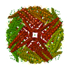
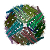
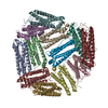
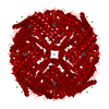
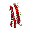
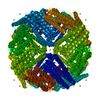
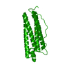
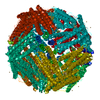
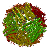
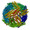
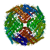
| Title | Structure of a modified mouse H-chain ferritin with a lanthanide binding motif | ||||||
 Components Components | Ferritin heavy chain | ||||||
 Keywords Keywords | METAL TRANSPORT / ferritin / lanthanide | ||||||
| Function / homology |  Function and homology information Function and homology informationIron uptake and transport / Golgi Associated Vesicle Biogenesis / iron ion sequestering activity / autolysosome / ferroxidase / intracellular sequestering of iron ion / ferroxidase activity / negative regulation of fibroblast proliferation / endocytic vesicle lumen / Neutrophil degranulation ...Iron uptake and transport / Golgi Associated Vesicle Biogenesis / iron ion sequestering activity / autolysosome / ferroxidase / intracellular sequestering of iron ion / ferroxidase activity / negative regulation of fibroblast proliferation / endocytic vesicle lumen / Neutrophil degranulation / ferric iron binding / ferrous iron binding / iron ion transport / immune response / iron ion binding / negative regulation of cell population proliferation / mitochondrion / extracellular region / identical protein binding / cytoplasmSimilarity search - Function | ||||||
| Biological species |  Mus musculus (house mouse) Mus musculus (house mouse) | ||||||
| Method | X-RAY DIFFRACTION / SYNCHROTRON / MOLECULAR REPLACEMENT / Resolution: 2.85 Å | ||||||
 Authors Authors | Baiocco, P. / Trabuco, M.C. | ||||||
| Funding support |  Italy, 1items Italy, 1items
| ||||||
 Citation Citation | Journal: PLoS ONE / Year: 2018 Title: Engineered ferritin for lanthanide binding. Authors: Calisti, L. / Trabuco, M.C. / Boffi, A. / Testi, C. / Montemiglio, L.C. / des Georges, A. / Benni, I. / Ilari, A. / Taciak, B. / Bialasek, M. / Rygiel, T. / Krol, M. / Baiocco, P. / Bonamore, A. | ||||||
| History |
|
- Structure visualization
| Structure viewer | Molecule: MolmilJmol/JSmol |
|---|
- Downloads & links
-Download
| PDBx/mmCIF format | 5oba.cif.gz | 807.9 KB | Display | PDBx/mmCIF format |
|---|---|---|---|---|
| PDB format | pdb5oba.ent.gz | 675.8 KB | Display | PDB format |
| PDBx/mmJSON format | 5oba.json.gz | Tree view | PDBx/mmJSON format | |
| Others |  Other downloads Other downloads |
-Validation report
| Arichive directory | https://data.pdbj.org/pub/pdb/validation_reports/ob/5obaftp://data.pdbj.org/pub/pdb/validation_reports/ob/5oba | HTTPS FTP |
|---|
-Related structure data
| Related structure data |  5obbC 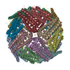 3wnwS S: Starting model for refinement C: citing same article ( |
|---|---|
| Similar structure data |


-Links
 PDBj
PDBj


- Assembly
| Deposited unit | 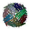
| ||||||||
|---|---|---|---|---|---|---|---|---|---|
| 1 |
| ||||||||
| Unit cell |
|
-Components
| #1: Protein | / Ferritin H subunit Mass: 22692.334 Da / Num. of mol.: 24 / Mutation: H177G Source method: isolated from a genetically manipulated source Source: (gene. exp.) Mus musculus (house mouse) / Gene: Fth1, Fth / Production host:  Escherichia coli (E. coli) / References: UniProt: P09528, ferroxidase Escherichia coli (E. coli) / References: UniProt: P09528, ferroxidase#2: Chemical | ChemComp-FE / Iron  Mass: 55.845 Da / Num. of mol.: 32 / Source method: obtained synthetically / Formula: Fe Mass: 55.845 Da / Num. of mol.: 32 / Source method: obtained synthetically / Formula: Fe#3: Water | ChemComp-HOH / | Water Mass: 18.015 Da / Num. of mol.: 12 / Source method: isolated from a natural source / Formula: H2O Mass: 18.015 Da / Num. of mol.: 12 / Source method: isolated from a natural source / Formula: H2O |
|---|
-Experimental details
-Experiment
| Experiment | Method: X-RAY DIFFRACTION / Number of used crystals: 1 |
|---|
- Sample preparation
| Crystal | Density Matthews: 3.49 Å3/Da / Density % sol: 64.74 % |
|---|---|
| Crystal grow | Temperature: 294 K / Method: vapor diffusion, hanging drop / pH: 8.5 / Details: Ammonium sulphate, Tris-HCl |
-Data collection
| Diffraction | Mean temperature: 100 K | ||||||||||||||||||||
|---|---|---|---|---|---|---|---|---|---|---|---|---|---|---|---|---|---|---|---|---|---|
| Diffraction source | Source: SYNCHROTRON / Site: ELETTRA / Beamline: 5.2R / Wavelength: 1 Å | ||||||||||||||||||||
| Detector | Type: DECTRIS PILATUS 2M / Detector: PIXEL / Date: Oct 11, 2016 | ||||||||||||||||||||
| Radiation | Protocol: SINGLE WAVELENGTH / Monochromatic (M) / Laue (L): M / Scattering type: x-ray | ||||||||||||||||||||
| Radiation wavelength | Wavelength: 1 Å / Relative weight: 1 | ||||||||||||||||||||
| Reflection twin |
| ||||||||||||||||||||
| Reflection | Resolution: 2.85→48.48 Å / Num. obs: 147573 / % possible obs: 100 % / Redundancy: 6.9 % / Net I/σ(I): 17.6 | ||||||||||||||||||||
| Reflection shell | Resolution: 2.85→2.9 Å / Redundancy: 6.8 % / Mean I/σ(I) obs: 2.5 / Num. unique obs: 4475 / % possible all: 100 |
- Processing
| Software |
| ||||||||||||||||||||||||||||||||||||||||||||||||||||||||||||
|---|---|---|---|---|---|---|---|---|---|---|---|---|---|---|---|---|---|---|---|---|---|---|---|---|---|---|---|---|---|---|---|---|---|---|---|---|---|---|---|---|---|---|---|---|---|---|---|---|---|---|---|---|---|---|---|---|---|---|---|---|---|
| Refinement | Method to determine structure: MOLECULAR REPLACEMENT Starting model: 3WNW Resolution: 2.85→48.48 Å / Cor.coef. Fo:Fc: 0.953 / Cor.coef. Fo:Fc free: 0.924 / SU B: 7.586 / SU ML: 0.145 / Cross valid method: THROUGHOUT / σ(F): 0 / ESU R: 0.145 / ESU R Free: 0.05 Details: HYDROGENS HAVE BEEN ADDED IN THE RIDING POSITIONS U VALUES : REFINED INDIVIDUALLY
| ||||||||||||||||||||||||||||||||||||||||||||||||||||||||||||
| Solvent computation | Ion probe radii: 0.8 Å / Shrinkage radii: 0.8 Å / VDW probe radii: 1.2 Å | ||||||||||||||||||||||||||||||||||||||||||||||||||||||||||||
| Displacement parameters | Biso max: 109.77 Å2 / Biso mean: 58.42 Å2 / Biso min: 13.97 Å2
| ||||||||||||||||||||||||||||||||||||||||||||||||||||||||||||
| Refinement step | Cycle: final / Resolution: 2.85→48.48 Å
| ||||||||||||||||||||||||||||||||||||||||||||||||||||||||||||
| Refine LS restraints |
| ||||||||||||||||||||||||||||||||||||||||||||||||||||||||||||
| LS refinement shell | Resolution: 2.848→2.921 Å / Rfactor Rfree error: 0 / Total num. of bins used: 20
|
