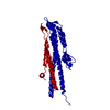
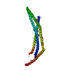
 Movie
Movie Controller


+ Open data

- Basic information
| Entry | Database: PDB / ID: 7mlw | ||||||
|---|---|---|---|---|---|---|---|
| Title | Burkholderia sp. TJI49 Guanidine-I riboswitch | ||||||
 Components Components | Guanidine-I riboswitch | ||||||
 Keywords Keywords |  RNA / Riboswitch / guanidine / potassium / ion / A-minor RNA / Riboswitch / guanidine / potassium / ion / A-minor | ||||||
| Function / homology | GUANIDINE / : / PHOSPHATE ION / STRONTIUM ION / RNA / RNA (> 10) / RNA (> 100) Function and homology information Function and homology information | ||||||
| Biological species |  Burkholderia sp. TJI49 (bacteria) Burkholderia sp. TJI49 (bacteria) | ||||||
| Method | X-RAY DIFFRACTION / SYNCHROTRON / MOLECULAR REPLACEMENT / Resolution: 2.7 Å | ||||||
 Authors Authors | Trachman, R.J. / Ferre-D'Amare, A.R. | ||||||
 Citation Citation | Journal: Rna / Year: 2021 Title: An uncommon [K + (Mg 2+ ) 2 ] metal ion triad imparts stability and selectivity to the Guanidine-I riboswitch. Authors: Trachman 3rd, R.J. / Ferre-D'Amare, A.R. | ||||||
| History |
|
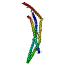
- Structure visualization
| Structure viewer | Molecule: MolmilJmol/JSmol |
|---|
- Downloads & links
-Download
| PDBx/mmCIF format | 7mlw.cif.gz | 182.1 KB | Display | PDBx/mmCIF format |
|---|---|---|---|---|
| PDB format | pdb7mlw.ent.gz | 120.5 KB | Display | PDB format |
| PDBx/mmJSON format | 7mlw.json.gz | Tree view | PDBx/mmJSON format | |
| Others |  Other downloads Other downloads |
-Validation report
| Arichive directory | https://data.pdbj.org/pub/pdb/validation_reports/ml/7mlwftp://data.pdbj.org/pub/pdb/validation_reports/ml/7mlw | HTTPS FTP |
|---|
-Related structure data
| Related structure data | 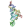 5t83S S: Starting model for refinement |
|---|---|
| Similar structure data |


-Links
 PDBj
PDBj


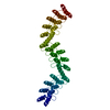
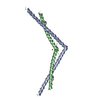
- Assembly
| Deposited unit | 
| ||||||||||||
|---|---|---|---|---|---|---|---|---|---|---|---|---|---|
| 1 |
| ||||||||||||
| Unit cell |
|
-Components
-RNA chain , 1 types, 1 molecules F
| #1: RNA chain | Mass: 41465.645 Da / Num. of mol.: 1 Source method: isolated from a genetically manipulated source Source: (gene. exp.) Burkholderia sp. TJI49 (bacteria)Production host: in vitro transcription vector pT7-TP(deltai) (others) |
|---|
-Non-polymers , 6 types, 26 molecules 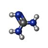


| #2: Chemical | ChemComp-GAI / Guanidine Mass: 59.070 Da / Num. of mol.: 1 / Source method: isolated from a natural source / Formula: CH5N3 / Feature type: SUBJECT OF INVESTIGATION Mass: 59.070 Da / Num. of mol.: 1 / Source method: isolated from a natural source / Formula: CH5N3 / Feature type: SUBJECT OF INVESTIGATION | ||||||||
|---|---|---|---|---|---|---|---|---|---|
| #3: Chemical | ChemComp-PO4 / Phosphate Mass: 94.971 Da / Num. of mol.: 4 / Source method: obtained synthetically / Formula: PO4 Mass: 94.971 Da / Num. of mol.: 4 / Source method: obtained synthetically / Formula: PO4#4: Chemical | Strontium Mass: 87.620 Da / Num. of mol.: 2 / Source method: obtained synthetically / Formula: Sr Mass: 87.620 Da / Num. of mol.: 2 / Source method: obtained synthetically / Formula: Sr#5: Chemical |  Mass: 24.305 Da / Num. of mol.: 3 / Source method: obtained synthetically / Formula: Mg / Feature type: SUBJECT OF INVESTIGATION Mass: 24.305 Da / Num. of mol.: 3 / Source method: obtained synthetically / Formula: Mg / Feature type: SUBJECT OF INVESTIGATION#6: Chemical |  Mass: 39.098 Da / Num. of mol.: 3 / Source method: obtained synthetically / Formula: K / Feature type: SUBJECT OF INVESTIGATION Mass: 39.098 Da / Num. of mol.: 3 / Source method: obtained synthetically / Formula: K / Feature type: SUBJECT OF INVESTIGATION#7: Water | ChemComp-HOH / | WaterMass: 18.015 Da / Num. of mol.: 13 / Source method: isolated from a natural source / Formula: H2O |
-Details
| Has ligand of interest | Y |
|---|
-Experimental details
-Experiment
| Experiment | Method: X-RAY DIFFRACTION / Number of used crystals: 1 |
|---|
- Sample preparation
| Crystal | Density Matthews: 2.48 Å3/Da / Density % sol: 50.44 % |
|---|---|
| Crystal grow | Temperature: 288 K / Method: vapor diffusion, sitting drop / pH: 7.5 Details: 50 mM HEPES (pH 7.5), 200 mM ammonium sulfate, 10 mM strontium chloride, 30% PEG 3350, 3% isopropanol |
-Data collection
| Diffraction | Mean temperature: 100 K / Serial crystal experiment: N |
|---|---|
| Diffraction source | Source: SYNCHROTRON / Site: ALS  / Beamline: 5.0.1 / Wavelength: 0.9919 Å / Beamline: 5.0.1 / Wavelength: 0.9919 Å |
| Detector | Type: DECTRIS PILATUS 6M / Detector: PIXEL / Date: Apr 14, 2017 |
| Radiation | Protocol: SINGLE WAVELENGTH / Monochromatic (M) / Laue (L): M / Scattering type: x-ray |
| Radiation wavelength | Wavelength: 0.9919 Å / Relative weight: 1 |
| Reflection | Resolution: 2.7→7 Å / Num. obs: 11350 / % possible obs: 98 % / Redundancy: 18.2 % / Biso Wilson estimate: 67.71 Å2 / CC1/2: 0.99 / Rmerge(I) obs: 0.07 / Net I/σ(I): 25.7 |
| Reflection shell | Resolution: 2.7→2.85 Å / Redundancy: 18.2 % / Rmerge(I) obs: 0.73 / Mean I/σ(I) obs: 4.1 / Num. unique obs: 936 / CC1/2: 0.92 / % possible all: 99.6 |
- Processing
| Software |
| |||||||||||||||||||||||||||||||||||||||||||||||||||||||||||||||
|---|---|---|---|---|---|---|---|---|---|---|---|---|---|---|---|---|---|---|---|---|---|---|---|---|---|---|---|---|---|---|---|---|---|---|---|---|---|---|---|---|---|---|---|---|---|---|---|---|---|---|---|---|---|---|---|---|---|---|---|---|---|---|---|---|
| Refinement | Method to determine structure: MOLECULAR REPLACEMENT Starting model: 5T83 Resolution: 2.7→6.99 Å / SU ML: 0.3528 / Cross valid method: FREE R-VALUE / σ(F): 1.36 / Phase error: 28.8037 Stereochemistry target values: GeoStd + Monomer Library + CDL v1.2
| |||||||||||||||||||||||||||||||||||||||||||||||||||||||||||||||
| Solvent computation | Shrinkage radii: 0.9 Å / VDW probe radii: 1.11 Å / Solvent model: FLAT BULK SOLVENT MODEL | |||||||||||||||||||||||||||||||||||||||||||||||||||||||||||||||
| Displacement parameters | Biso mean: 98.08 Å2 | |||||||||||||||||||||||||||||||||||||||||||||||||||||||||||||||
| Refinement step | Cycle: LAST / Resolution: 2.7→6.99 Å
| |||||||||||||||||||||||||||||||||||||||||||||||||||||||||||||||
| Refine LS restraints |
| |||||||||||||||||||||||||||||||||||||||||||||||||||||||||||||||
| LS refinement shell |
| |||||||||||||||||||||||||||||||||||||||||||||||||||||||||||||||
| Refinement TLS params. | Method: refined / Origin x: -13.8534670905 Å / Origin y: 3.73034604913 Å / Origin z: -18.2165343402 Å
| |||||||||||||||||||||||||||||||||||||||||||||||||||||||||||||||
| Refinement TLS group | Selection details: all |
