 Movie
Movie Controller


+ Open data

- Basic information
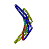
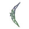
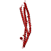
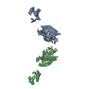
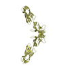
| Entry | Database: PDB / ID: 2q12 | ||||||
|---|---|---|---|---|---|---|---|
| Title | Crystal Structure of BAR domain of APPL1 | ||||||
 Components Components | DCC-interacting protein 13 alpha | ||||||
 Keywords Keywords |  PROTEIN TRANSPORT / APPL1 / BAR domain PROTEIN TRANSPORT / APPL1 / BAR domain | ||||||
| Function / homology |  Function and homology information Function and homology informationnegative regulation of Fc-gamma receptor signaling pathway involved in phagocytosis / positive regulation of macropinocytosis / adiponectin-activated signaling pathway / macropinosome / regulation of fibroblast migration / regulation of glucose import / maintenance of synapse structure / protein kinase B binding / signaling / positive regulation of melanin biosynthetic process ...negative regulation of Fc-gamma receptor signaling pathway involved in phagocytosis / positive regulation of macropinocytosis / adiponectin-activated signaling pathway / macropinosome / regulation of fibroblast migration / regulation of glucose import / maintenance of synapse structure / protein kinase B binding / signaling / positive regulation of melanin biosynthetic process / regulation of toll-like receptor 4 signaling pathway / vesicle membrane / positive regulation of cytokine production involved in inflammatory response / early phagosome / Caspase activation via Dependence Receptors in the absence of ligand / cellular response to hepatocyte growth factor stimulus / intracellular vesicle / phosphatidylserine binding / beta-tubulin binding / regulation of innate immune response / regulation of G1/S transition of mitotic cell cycle / regulation of protein localization to plasma membrane / ruffle / phosphatidylinositol binding / transforming growth factor beta receptor signaling pathway / positive regulation of glucose import / protein import into nucleus / presynapse / insulin receptor signaling pathway / cytoplasmic vesicle / early endosome membrane / postsynapse / early endosome / endosome membrane / endosome / cell cycle / glutamatergic synapse / protein-containing complex binding / signal transduction / protein homodimerization activity / extracellular exosome / membrane / identical protein binding / nucleus / plasma membrane / cytosol / cytoplasmSimilarity search - Function | ||||||
| Biological species |  Homo sapiens (human) Homo sapiens (human) | ||||||
| Method | X-RAY DIFFRACTION / SYNCHROTRON / SAD / Resolution: 1.79 Å | ||||||
 Authors Authors | Zhang, X.C. / Zhu, G. | ||||||
 Citation Citation | Journal: Embo J. / Year: 2007 Title: Structure of the APPL1 BAR-PH domain and characterization of its interaction with Rab5. Authors: Zhu, G. / Chen, J. / Liu, J. / Brunzelle, J.S. / Huang, B. / Wakeham, N. / Terzyan, S. / Li, X. / Rao, Z. / Li, G. / Zhang, X.C. | ||||||
| History |
|
- Structure visualization
| Structure viewer | Molecule: MolmilJmol/JSmol |
|---|
- Downloads & links
-Download
| PDBx/mmCIF format | 2q12.cif.gz | 61.1 KB | Display | PDBx/mmCIF format |
|---|---|---|---|---|
| PDB format | pdb2q12.ent.gz | 49.1 KB | Display | PDB format |
| PDBx/mmJSON format | 2q12.json.gz | Tree view | PDBx/mmJSON format | |
| Others |  Other downloads Other downloads |
-Validation report
| Arichive directory | https://data.pdbj.org/pub/pdb/validation_reports/q1/2q12ftp://data.pdbj.org/pub/pdb/validation_reports/q1/2q12 | HTTPS FTP |
|---|
-Related structure data


-Links
 PDBj
PDBj


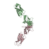
- Assembly
| Deposited unit | 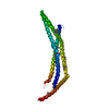
| ||||||||
|---|---|---|---|---|---|---|---|---|---|
| 1 | 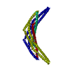
| ||||||||
| Unit cell |
| ||||||||
| Details | The biological dimer is generated from molecules in asymmetric unit and operation -x+1, -y, z |
-Components
| #1: Protein | Mass: 31157.240 Da / Num. of mol.: 1 / Fragment: residues 5-265, BAR domain Source method: isolated from a genetically manipulated source Source: (gene. exp.) Homo sapiens (human) / Gene: APPL1, APPL, DIP13A, KIAA1428 / Plasmid: pET28a / Production host:  Escherichia coli (E. coli) / Strain (production host): B834 / References: UniProt: Q9UKG1 Escherichia coli (E. coli) / Strain (production host): B834 / References: UniProt: Q9UKG1 |
|---|---|
| #2: Water | ChemComp-HOH / Water Mass: 18.015 Da / Num. of mol.: 147 / Source method: isolated from a natural source / Formula: H2O Mass: 18.015 Da / Num. of mol.: 147 / Source method: isolated from a natural source / Formula: H2O |
-Experimental details
-Experiment
| Experiment | Method: X-RAY DIFFRACTION / Number of used crystals: 1 |
|---|
- Sample preparation
| Crystal | Density Matthews: 2.02 Å3/Da / Density % sol: 39.22 % |
|---|---|
| Crystal grow | Temperature: 293 K / Method: vapor diffusion, hanging drop / pH: 8 Details: 0.1 M magnesium formate, pH 8.0, VAPOR DIFFUSION, HANGING DROP, temperature 293K |
-Data collection
| Diffraction | Mean temperature: 100 K |
|---|---|
| Diffraction source | Source: SYNCHROTRON / Site: APS  / Beamline: 22-BM / Wavelength: 0.9793 Å / Beamline: 22-BM / Wavelength: 0.9793 Å |
| Detector | Type: MARMOSAIC 225 mm CCD / Detector: CCD / Date: Mar 16, 2006 |
| Radiation | Monochromator: Rosenbaum-Rock double-crystal monochromator / Protocol: SINGLE WAVELENGTH / Monochromatic (M) / Laue (L): M / Scattering type: x-ray |
| Radiation wavelength | Wavelength: 0.9793 Å / Relative weight: 1 |
| Reflection | Resolution: 1.79→50 Å / Num. all: 23548 / Num. obs: 23518 / % possible obs: 95.7 % / Observed criterion σ(F): -6 / Observed criterion σ(I): -3 / Redundancy: 3.2 % / Biso Wilson estimate: 25.8 Å2 / Rmerge(I) obs: 0.081 / Net I/σ(I): 10.1 |
| Reflection shell | Resolution: 1.8→1.86 Å / Redundancy: 3 % / Rmerge(I) obs: 0.44 / Mean I/σ(I) obs: 2.19 / Num. unique all: 1897 / % possible all: 78.1 |
- Processing
| Software |
| ||||||||||||||||||||||||||||||||||||||||||||||||||||||||||||||||||||||||||||||||||||||||||
|---|---|---|---|---|---|---|---|---|---|---|---|---|---|---|---|---|---|---|---|---|---|---|---|---|---|---|---|---|---|---|---|---|---|---|---|---|---|---|---|---|---|---|---|---|---|---|---|---|---|---|---|---|---|---|---|---|---|---|---|---|---|---|---|---|---|---|---|---|---|---|---|---|---|---|---|---|---|---|---|---|---|---|---|---|---|---|---|---|---|---|---|
| Refinement | Method to determine structure: SAD / Resolution: 1.79→50 Å / Cor.coef. Fo:Fc: 0.95 / Cor.coef. Fo:Fc free: 0.935 / SU B: 3.355 / SU ML: 0.106 / Isotropic thermal model: Isotropic / Cross valid method: THROUGHOUT / σ(F): 0 / ESU R: 0.155 / ESU R Free: 0.146 / Stereochemistry target values: MAXIMUM LIKELIHOOD
| ||||||||||||||||||||||||||||||||||||||||||||||||||||||||||||||||||||||||||||||||||||||||||
| Solvent computation | Ion probe radii: 0.8 Å / Shrinkage radii: 0.8 Å / VDW probe radii: 1.4 Å / Solvent model: MASK | ||||||||||||||||||||||||||||||||||||||||||||||||||||||||||||||||||||||||||||||||||||||||||
| Displacement parameters | Biso mean: 34.596 Å2
| ||||||||||||||||||||||||||||||||||||||||||||||||||||||||||||||||||||||||||||||||||||||||||
| Refinement step | Cycle: LAST / Resolution: 1.79→50 Å
| ||||||||||||||||||||||||||||||||||||||||||||||||||||||||||||||||||||||||||||||||||||||||||
| Refine LS restraints |
| ||||||||||||||||||||||||||||||||||||||||||||||||||||||||||||||||||||||||||||||||||||||||||
| LS refinement shell | Resolution: 1.792→1.839 Å / Total num. of bins used: 20
|
