 Movie
Movie Controller

[English] 日本語
 Yorodumi
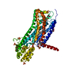
- PDB-7m8w: XFEL crystal structure of the prostaglandin D2 receptor CRTH2 in ... -
+ Open data

- Basic information
| Entry | Database: PDB / ID: 7m8w | ||||||
|---|---|---|---|---|---|---|---|
| Title | XFEL crystal structure of the prostaglandin D2 receptor CRTH2 in complex with 15R-methyl-PGD2 | ||||||
 Components Components | Prostaglandin D2 receptor 2, Endolysin chimera | ||||||
 Keywords Keywords | MEMBRANE PROTEIN / prostaglandin D2 receptor / CRTH2 / 15R-methyl-PGD2 / G protein-coupled receptor / GPCR / Endolysin fusion / LCP | ||||||
| Function / homology |  Function and homology information Function and homology informationprostaglandin J receptor activity / prostaglandin D receptor activity / prostaglandin F receptor activity / Prostanoid ligand receptors / negative regulation of male germ cell proliferation / positive regulation of G protein-coupled receptor signaling pathway / neuropeptide binding / neuropeptide signaling pathway / viral release from host cell by cytolysis / adenylate cyclase-inhibiting G protein-coupled receptor signaling pathway ...prostaglandin J receptor activity / prostaglandin D receptor activity / prostaglandin F receptor activity / Prostanoid ligand receptors / negative regulation of male germ cell proliferation / positive regulation of G protein-coupled receptor signaling pathway / neuropeptide binding / neuropeptide signaling pathway / viral release from host cell by cytolysis / adenylate cyclase-inhibiting G protein-coupled receptor signaling pathway / peptidoglycan catabolic process / G protein-coupled receptor activity / calcium-mediated signaling / chemotaxis / cell wall macromolecule catabolic process / lysozyme / lysozyme activity / G alpha (i) signalling events / host cell cytoplasm / defense response to bacterium / neuron projection / immune response / G protein-coupled receptor signaling pathway / plasma membraneSimilarity search - Function | ||||||
| Biological species |  Homo sapiens (human) Homo sapiens (human) Enterobacteria phage T4 (virus) Enterobacteria phage T4 (virus) | ||||||
| Method | X-RAY DIFFRACTION / FREE ELECTRON LASER / MOLECULAR REPLACEMENT / Resolution: 2.61 Å | ||||||
 Authors Authors | Shiriaeva, A. / Han, G.W. / Cherezov, V. | ||||||
 Citation Citation | Journal: Proc.Natl.Acad.Sci.USA / Year: 2021 Title: Molecular basis for lipid recognition by the prostaglandin D 2 receptor CRTH2. Authors: Liu, H. / Deepak, R.N.V.K. / Shiriaeva, A. / Gati, C. / Batyuk, A. / Hu, H. / Weierstall, U. / Liu, W. / Wang, L. / Cherezov, V. / Fan, H. / Zhang, C. | ||||||
| History |
|
- Structure visualization
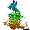
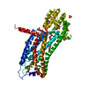
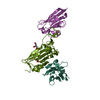
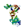
| Structure viewer | Molecule: MolmilJmol/JSmol |
|---|
- Downloads & links
-Download
| PDBx/mmCIF format | 7m8w.cif.gz | 193.8 KB | Display | PDBx/mmCIF format |
|---|---|---|---|---|
| PDB format | pdb7m8w.ent.gz | 150.9 KB | Display | PDB format |
| PDBx/mmJSON format | 7m8w.json.gz | Tree view | PDBx/mmJSON format | |
| Others |  Other downloads Other downloads |
-Validation report
| Arichive directory | https://data.pdbj.org/pub/pdb/validation_reports/m8/7m8wftp://data.pdbj.org/pub/pdb/validation_reports/m8/7m8w | HTTPS FTP |
|---|
-Related structure data
| Related structure data |  6d27S S: Starting model for refinement |
|---|---|
| Similar structure data |


-Links
 PDBj
PDBj


- Assembly
| Deposited unit | 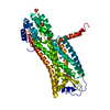
| ||||||||
|---|---|---|---|---|---|---|---|---|---|
| 1 |
| ||||||||
| Unit cell |
| ||||||||
| Details | AUTHORS STATE THAT THE BIOLOGICAL UNIT IS UNKNOWN. |
-Components
-Protein , 1 types, 1 molecules A
| #1: Protein | Mass: 52228.727 Da / Num. of mol.: 1 Source method: isolated from a genetically manipulated source Source: (gene. exp.) Homo sapiens (human), (gene. exp.) Enterobacteria phage T4 (virus)Gene: PTGDR2, CRTH2, DL1R, GPR44, e, T4Tp126 / Production host:   Spodoptera frugiperda (fall armyworm) / References: UniProt: Q9Y5Y4, UniProt: D9IEF7 Spodoptera frugiperda (fall armyworm) / References: UniProt: Q9Y5Y4, UniProt: D9IEF7 |
|---|
-Non-polymers , 5 types, 20 molecules 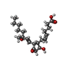


| #2: Chemical | ChemComp-YSS /  Mass: 366.492 Da / Num. of mol.: 1 / Source method: obtained synthetically / Formula: C21H34O5 / Feature type: SUBJECT OF INVESTIGATION Mass: 366.492 Da / Num. of mol.: 1 / Source method: obtained synthetically / Formula: C21H34O5 / Feature type: SUBJECT OF INVESTIGATION | ||
|---|---|---|---|
| #3: Chemical | ChemComp-NA /  Mass: 22.990 Da / Num. of mol.: 1 / Source method: obtained synthetically / Formula: Na Mass: 22.990 Da / Num. of mol.: 1 / Source method: obtained synthetically / Formula: Na | ||
| #4: Chemical | ChemComp-FLC / Citric acid Mass: 189.100 Da / Num. of mol.: 1 / Source method: obtained synthetically / Formula: C6H5O7 Mass: 189.100 Da / Num. of mol.: 1 / Source method: obtained synthetically / Formula: C6H5O7 | ||
| #5: Chemical | ChemComp-SO4 / Sulfate Mass: 96.063 Da / Num. of mol.: 5 / Source method: obtained synthetically / Formula: SO4 Mass: 96.063 Da / Num. of mol.: 5 / Source method: obtained synthetically / Formula: SO4#6: Water | ChemComp-HOH / | WaterMass: 18.015 Da / Num. of mol.: 12 / Source method: isolated from a natural source / Formula: H2O |
-Details
| Has ligand of interest | Y |
|---|
-Experimental details
-Experiment
| Experiment | Method: X-RAY DIFFRACTION / Number of used crystals: 20358 |
|---|
- Sample preparation
| Crystal | Density Matthews: 4.43 Å3/Da / Density % sol: 72.22 % |
|---|---|
| Crystal grow | Temperature: 283 K / Method: lipidic cubic phase / pH: 6 Details: sodium chloride, lithium sulphate, sodium citrate tribasic dihydrate, PEG 300, propylene glycol P400, PGD2 |
-Data collection
| Diffraction | Mean temperature: 293 K / Serial crystal experiment: Y |
|---|---|
| Diffraction source | Source: FREE ELECTRON LASER / Site: SLAC LCLS  / Beamline: CXI / Wavelength: 1.33 Å / Beamline: CXI / Wavelength: 1.33 Å |
| Detector | Type: CS-PAD CXI-1 / Detector: PIXEL / Date: Nov 4, 2018 / Frequency: 120 |
| Radiation | Protocol: SINGLE WAVELENGTH / Monochromatic (M) / Laue (L): M / Scattering type: x-ray |
| Radiation wavelength | Wavelength: 1.33 Å / Relative weight: 1 |
| Reflection | Resolution: 2.6→32.43 Å / Num. obs: 29269 / % possible obs: 98.9 % / Redundancy: 61.8 % / CC star: 0.99 / R split: 0.264 / Net I/σ(I): 2.4 |
| Reflection shell | Resolution: 2.6→2.69 Å / Redundancy: 4.9 % / Mean I/σ(I) obs: 0.2 / Num. unique obs: 12462 / CC star: 0.512 / R split: 5.14 / % possible all: 90.6 |
| Serial crystallography measurement | Collection time total: 2 hours / Collimation: KB mirrors / Focal spot size: 2 µm2 / Pulse duration: 35 fsec. / Pulse energy: 0.1 µJ / Pulse photon energy: 9.5 keV / XFEL pulse repetition rate: 120 Hz |
| Serial crystallography sample delivery | Method: injection |
| Serial crystallography sample delivery injection | Carrier solvent: LCP / Description: LCP injector / Flow rate: 0.44 µL/min / Injector diameter: 50 µm / Injector temperature: 293 K / Jet diameter: 50 µm / Power by: HPLC pump |
| Serial crystallography data reduction | Crystal hits: 20358 / Frames total: 850000 |
- Processing
| Software |
| |||||||||||||||||||||||||||||||||||||||||||||||||||||||||||||||||||||||||||||||||||||||||||||||||||||||||||||||||||||||||||||
|---|---|---|---|---|---|---|---|---|---|---|---|---|---|---|---|---|---|---|---|---|---|---|---|---|---|---|---|---|---|---|---|---|---|---|---|---|---|---|---|---|---|---|---|---|---|---|---|---|---|---|---|---|---|---|---|---|---|---|---|---|---|---|---|---|---|---|---|---|---|---|---|---|---|---|---|---|---|---|---|---|---|---|---|---|---|---|---|---|---|---|---|---|---|---|---|---|---|---|---|---|---|---|---|---|---|---|---|---|---|---|---|---|---|---|---|---|---|---|---|---|---|---|---|---|---|---|
| Refinement | Method to determine structure: MOLECULAR REPLACEMENT Starting model: 6D27 Resolution: 2.61→32.43 Å / SU ML: 0.33 / Cross valid method: THROUGHOUT / σ(F): 1.33 / Phase error: 32.26 / Stereochemistry target values: ML
| |||||||||||||||||||||||||||||||||||||||||||||||||||||||||||||||||||||||||||||||||||||||||||||||||||||||||||||||||||||||||||||
| Solvent computation | Shrinkage radii: 0.9 Å / VDW probe radii: 1.11 Å / Solvent model: FLAT BULK SOLVENT MODEL | |||||||||||||||||||||||||||||||||||||||||||||||||||||||||||||||||||||||||||||||||||||||||||||||||||||||||||||||||||||||||||||
| Displacement parameters | Biso max: 155.54 Å2 / Biso mean: 52.3671 Å2 / Biso min: 10.52 Å2 | |||||||||||||||||||||||||||||||||||||||||||||||||||||||||||||||||||||||||||||||||||||||||||||||||||||||||||||||||||||||||||||
| Refinement step | Cycle: final / Resolution: 2.61→32.43 Å
| |||||||||||||||||||||||||||||||||||||||||||||||||||||||||||||||||||||||||||||||||||||||||||||||||||||||||||||||||||||||||||||
| LS refinement shell | Refine-ID: X-RAY DIFFRACTION / Rfactor Rfree error: 0 / Total num. of bins used: 6
| |||||||||||||||||||||||||||||||||||||||||||||||||||||||||||||||||||||||||||||||||||||||||||||||||||||||||||||||||||||||||||||
| Refinement TLS params. | Method: refined / Refine-ID: X-RAY DIFFRACTION
| |||||||||||||||||||||||||||||||||||||||||||||||||||||||||||||||||||||||||||||||||||||||||||||||||||||||||||||||||||||||||||||
| Refinement TLS group |
|
