 Movie
Movie Controller


+ Open data

- Basic information
| Entry | Database: PDB / ID: 7ksb | ||||||
|---|---|---|---|---|---|---|---|
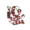
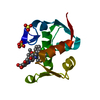
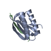
| Title | Crystal structure on Act c 10.0101 | ||||||
 Components Components | Non-specific lipid-transfer protein 1 | ||||||
 Keywords Keywords |  ALLERGEN / nsLTP / food allergen ALLERGEN / nsLTP / food allergen | ||||||
| Function / homology | Plant non-specific lipid-transfer protein/Par allergen / Protease inhibitor/seed storage/LTP family / Plant lipid transfer protein / seed storage protein / trypsin-alpha amylase inhibitor domain family / Bifunctional inhibitor/plant lipid transfer protein/seed storage helical domain / Bifunctional inhibitor/plant lipid transfer protein/seed storage helical domain superfamily / lipid transport / lipid binding / Non-specific lipid-transfer protein 1 Function and homology information Function and homology information | ||||||
| Biological species |  Actinidia chinensis var. chinensis (plant) Actinidia chinensis var. chinensis (plant) | ||||||
| Method | X-RAY DIFFRACTION / SYNCHROTRON / MOLECULAR REPLACEMENT / Resolution: 1.95 Å | ||||||
 Authors Authors | Pote, S. / O'Malley, A. / Gawlicka-Chruszcz, A. / Giangrieco, I. / Ciardiello, M.A. / Chruszcz, M. | ||||||
| Funding support |  United States, 1items United States, 1items
| ||||||
 Citation Citation | Journal: Molecules / Year: 2021 Title: Structural Characterization of Act c 10.0101 and Pun g 1.0101-Allergens from the Non-Specific Lipid Transfer Protein Family. Authors: O'Malley, A. / Pote, S. / Giangrieco, I. / Tuppo, L. / Gawlicka-Chruszcz, A. / Kowal, K. / Ciardiello, M.A. / Chruszcz, M. | ||||||
| History |
|
- Structure visualization
| Structure viewer | Molecule: MolmilJmol/JSmol |
|---|
- Downloads & links
-Download
| PDBx/mmCIF format | 7ksb.cif.gz | 80.3 KB | Display | PDBx/mmCIF format |
|---|---|---|---|---|
| PDB format | pdb7ksb.ent.gz | 60.1 KB | Display | PDB format |
| PDBx/mmJSON format | 7ksb.json.gz | Tree view | PDBx/mmJSON format | |
| Others |  Other downloads Other downloads |
-Validation report
| Arichive directory | https://data.pdbj.org/pub/pdb/validation_reports/ks/7ksbftp://data.pdbj.org/pub/pdb/validation_reports/ks/7ksb | HTTPS FTP |
|---|
-Related structure data
| Related structure data |  7kscC 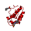 1rzlS S: Starting model for refinement C: citing same article ( |
|---|---|
| Similar structure data |


-Links
 PDBj
PDBj
- Assembly
| Deposited unit | 
| ||||||||||||||||||
|---|---|---|---|---|---|---|---|---|---|---|---|---|---|---|---|---|---|---|---|
| 1 | 
| ||||||||||||||||||
| 2 | 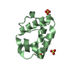
| ||||||||||||||||||
| 3 |
| ||||||||||||||||||
| Unit cell |
| ||||||||||||||||||
| Noncrystallographic symmetry (NCS) | NCS domain:
NCS domain segments: Component-ID: 0 / Ens-ID: 1 / Beg auth comp-ID: ALA / Beg label comp-ID: ALA / End auth comp-ID: SER / End label comp-ID: SER / Refine code: 0 / Auth seq-ID: 1 - 92 / Label seq-ID: 1 - 92
|
-Components
| #1: Protein | Mass: 9472.930 Da / Num. of mol.: 2 / Source method: isolated from a natural source Source: (natural) Actinidia chinensis var. chinensis (plant)References: UniProt: P85204 #2: Chemical | ChemComp-SO4 / Sulfate  Mass: 96.063 Da / Num. of mol.: 6 / Source method: obtained synthetically / Formula: SO4 Mass: 96.063 Da / Num. of mol.: 6 / Source method: obtained synthetically / Formula: SO4#3: Water | ChemComp-HOH / | Water Mass: 18.015 Da / Num. of mol.: 83 / Source method: isolated from a natural source / Formula: H2O Mass: 18.015 Da / Num. of mol.: 83 / Source method: isolated from a natural source / Formula: H2OHas ligand of interest | N | |
|---|
-Experimental details
-Experiment
| Experiment | Method: X-RAY DIFFRACTION / Number of used crystals: 1 |
|---|
- Sample preparation
| Crystal | Density Matthews: 1.87 Å3/Da / Density % sol: 34.16 % |
|---|---|
| Crystal grow | Temperature: 295 K / Method: vapor diffusion / pH: 4 Details: 0.1 M citric acid buffer, pH 4.0 with 3.0 M ammonium sulfate |
-Data collection
| Diffraction | Mean temperature: 100 K / Serial crystal experiment: N |
|---|---|
| Diffraction source | Source: SYNCHROTRON / Site: APS / Beamline: 22-ID / Wavelength: 1 Å |
| Detector | Type: RAYONIX MX300-HS / Detector: CCD / Date: Jun 20, 2017 |
| Radiation | Protocol: SINGLE WAVELENGTH / Monochromatic (M) / Laue (L): M / Scattering type: x-ray |
| Radiation wavelength | Wavelength: 1 Å / Relative weight: 1 |
| Reflection | Resolution: 1.95→40 Å / Num. obs: 10217 / % possible obs: 98.7 % / Redundancy: 4 % / Rpim(I) all: 0.057 / Rrim(I) all: 0.113 / Net I/σ(I): 25.1 |
| Reflection shell | Resolution: 1.95→1.98 Å / Redundancy: 3.9 % / Mean I/σ(I) obs: 4.3 / Num. unique obs: 497 / CC1/2: 0.904 / Rpim(I) all: 0.24 / Rrim(I) all: 0.483 / % possible all: 98.2 |
- Processing
| Software |
| |||||||||||||||||||||||||||||||||||||||||||||||||||||||||||||||||||||||||||||||||||||||||||||||||||||||||||||||||||||||||||||
|---|---|---|---|---|---|---|---|---|---|---|---|---|---|---|---|---|---|---|---|---|---|---|---|---|---|---|---|---|---|---|---|---|---|---|---|---|---|---|---|---|---|---|---|---|---|---|---|---|---|---|---|---|---|---|---|---|---|---|---|---|---|---|---|---|---|---|---|---|---|---|---|---|---|---|---|---|---|---|---|---|---|---|---|---|---|---|---|---|---|---|---|---|---|---|---|---|---|---|---|---|---|---|---|---|---|---|---|---|---|---|---|---|---|---|---|---|---|---|---|---|---|---|---|---|---|---|
| Refinement | Method to determine structure: MOLECULAR REPLACEMENT Starting model: 1RZL Resolution: 1.95→27.91 Å / Cor.coef. Fo:Fc: 0.946 / Cor.coef. Fo:Fc free: 0.91 / SU B: 10.241 / SU ML: 0.152 / Cross valid method: THROUGHOUT / σ(F): 0 / ESU R: 0.238 / ESU R Free: 0.197 / Stereochemistry target values: MAXIMUM LIKELIHOOD Details: HYDROGENS HAVE BEEN ADDED IN THE RIDING POSITIONS U VALUES : WITH TLS ADDED
| |||||||||||||||||||||||||||||||||||||||||||||||||||||||||||||||||||||||||||||||||||||||||||||||||||||||||||||||||||||||||||||
| Solvent computation | Ion probe radii: 0.8 Å / Shrinkage radii: 0.8 Å / VDW probe radii: 1.2 Å / Solvent model: MASK | |||||||||||||||||||||||||||||||||||||||||||||||||||||||||||||||||||||||||||||||||||||||||||||||||||||||||||||||||||||||||||||
| Displacement parameters | Biso max: 72.19 Å2 / Biso mean: 26.802 Å2 / Biso min: 15.24 Å2
| |||||||||||||||||||||||||||||||||||||||||||||||||||||||||||||||||||||||||||||||||||||||||||||||||||||||||||||||||||||||||||||
| Refinement step | Cycle: final / Resolution: 1.95→27.91 Å
| |||||||||||||||||||||||||||||||||||||||||||||||||||||||||||||||||||||||||||||||||||||||||||||||||||||||||||||||||||||||||||||
| Refine LS restraints |
| |||||||||||||||||||||||||||||||||||||||||||||||||||||||||||||||||||||||||||||||||||||||||||||||||||||||||||||||||||||||||||||
| Refine LS restraints NCS | Ens-ID: 1 / Number: 2528 / Refine-ID: X-RAY DIFFRACTION / Type: interatomic distance / Rms dev position: 0.11 Å / Weight position: 0.05
| |||||||||||||||||||||||||||||||||||||||||||||||||||||||||||||||||||||||||||||||||||||||||||||||||||||||||||||||||||||||||||||
| LS refinement shell | Resolution: 1.95→2.001 Å / Rfactor Rfree error: 0 / Total num. of bins used: 20
| |||||||||||||||||||||||||||||||||||||||||||||||||||||||||||||||||||||||||||||||||||||||||||||||||||||||||||||||||||||||||||||
| Refinement TLS params. | Method: refined / Refine-ID: X-RAY DIFFRACTION
| |||||||||||||||||||||||||||||||||||||||||||||||||||||||||||||||||||||||||||||||||||||||||||||||||||||||||||||||||||||||||||||
| Refinement TLS group |
|
