 Movie
Movie Controller

[English] 日本語
 Yorodumi
- PDB-4ht6: The Structure of a Yeast Dynein Dyn2-Pac11 Complex and Effect on ... -
+ Open data

- Basic information
| Entry | Database: PDB / ID: 4ht6 | ||||||
|---|---|---|---|---|---|---|---|
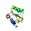
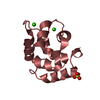
| Title | The Structure of a Yeast Dynein Dyn2-Pac11 Complex and Effect on Single Molecule Dynein Motor Activity | ||||||
 Components Components |
| ||||||
 Keywords Keywords |  MOTOR PROTEIN / Dimerization / Dynein / Intermediate Chain / Light Chain / Dynein Intermediate Chain Dynein Heavy Chain MOTOR PROTEIN / Dimerization / Dynein / Intermediate Chain / Light Chain / Dynein Intermediate Chain Dynein Heavy Chain | ||||||
| Function / homology |  Function and homology information Function and homology information: / peroxisomal importomer complex / dynein light chain binding / dynein heavy chain binding / establishment of mitotic spindle localization / nuclear migration along microtubule / nuclear pore complex assembly / cytoplasmic dynein complex / nucleocytoplasmic transport / dynein intermediate chain binding ...: / peroxisomal importomer complex / dynein light chain binding / dynein heavy chain binding / establishment of mitotic spindle localization / nuclear migration along microtubule / nuclear pore complex assembly / cytoplasmic dynein complex / nucleocytoplasmic transport / dynein intermediate chain binding / microtubule-based movement / establishment of mitotic spindle orientation / mRNA transport / cytoplasmic microtubule / nuclear pore / Neutrophil degranulation / nuclear periphery / protein transport / nuclear envelope / protein-containing complex binding / cytoplasmSimilarity search - Function | ||||||
| Biological species |  Saccharomyces cerevisiae (brewer's yeast) Saccharomyces cerevisiae (brewer's yeast) | ||||||
| Method | X-RAY DIFFRACTION / SYNCHROTRON / MOLECULAR REPLACEMENT / Resolution: 1.9 Å | ||||||
 Authors Authors | Slep, K.C. / Romes, E.R. | ||||||
 Citation Citation | Journal: Mol Biol Cell / Year: 2013 Title: The yeast dynein Dyn2-Pac11 complex is a dynein dimerization/processivity factor: structural and single-molecule characterization. Authors: Rao, L. / Romes, E.M. / Nicholas, M.P. / Brenner, S. / Tripathy, A. / Gennerich, A. / Slep, K.C. | ||||||
| History |
|
- Structure visualization
| Structure viewer | Molecule: MolmilJmol/JSmol |
|---|
- Downloads & links
-Download
| PDBx/mmCIF format | 4ht6.cif.gz | 137.9 KB | Display | PDBx/mmCIF format |
|---|---|---|---|---|
| PDB format | pdb4ht6.ent.gz | 109.2 KB | Display | PDB format |
| PDBx/mmJSON format | 4ht6.json.gz | Tree view | PDBx/mmJSON format | |
| Others |  Other downloads Other downloads |
-Validation report
| Arichive directory | https://data.pdbj.org/pub/pdb/validation_reports/ht/4ht6ftp://data.pdbj.org/pub/pdb/validation_reports/ht/4ht6 | HTTPS FTP |
|---|
-Related structure data
| Related structure data |  4ds1S S: Starting model for refinement |
|---|---|
| Similar structure data |


-Links
 PDBj
PDBj


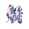
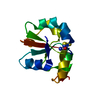
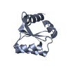
- Assembly
| Deposited unit | 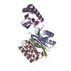
| ||||||||
|---|---|---|---|---|---|---|---|---|---|
| 1 | 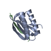
| ||||||||
| 2 | 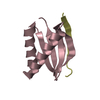
| ||||||||
| 3 | 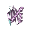
| ||||||||
| 4 | 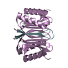
| ||||||||
| Unit cell |
| ||||||||
| Components on special symmetry positions |
|
-Components
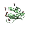
| #1: Protein | Mass: 10868.436 Da / Num. of mol.: 3 Source method: isolated from a genetically manipulated source Source: (gene. exp.) Saccharomyces cerevisiae (brewer's yeast)Strain: S288c / Gene: DYN2, SLC1, YDR424C / Plasmid: pGEX-6P2 / Production host:  Escherichia coli (E. coli) / Strain (production host): BL21 DE3 (pLysS) / References: UniProt: Q02647 Escherichia coli (E. coli) / Strain (production host): BL21 DE3 (pLysS) / References: UniProt: Q02647#2: Protein/peptide | Mass: 1282.377 Da / Num. of mol.: 3 / Fragment: residues 75-85 / Source method: obtained synthetically / Source: (synth.) Saccharomyces cerevisiae (brewer's yeast) / References: UniProt: P40960#3: Water | ChemComp-HOH / | Water Mass: 18.015 Da / Num. of mol.: 274 / Source method: isolated from a natural source / Formula: H2O Mass: 18.015 Da / Num. of mol.: 274 / Source method: isolated from a natural source / Formula: H2O |
|---|
-Experimental details
-Experiment
| Experiment | Method: X-RAY DIFFRACTION / Number of used crystals: 1 |
|---|
- Sample preparation
| Crystal | Density Matthews: 2.21 Å3/Da / Density % sol: 44.4 % |
|---|---|
| Crystal grow | Temperature: 293 K / Method: vapor diffusion, hanging drop / pH: 6.8 Details: 0.4M sodium phosphate monobasic, 0.1M 1,6-hexanediol, 25% PEG 3350 (w/v), pH 6.8, VAPOR DIFFUSION, HANGING DROP, temperature 293K |
-Data collection
| Diffraction | Mean temperature: 100 K | |||||||||||||||||||||||||||||||||
|---|---|---|---|---|---|---|---|---|---|---|---|---|---|---|---|---|---|---|---|---|---|---|---|---|---|---|---|---|---|---|---|---|---|---|
| Diffraction source | Source: SYNCHROTRON / Site: APS  / Beamline: 22-ID / Wavelength: 0.9792 Å / Beamline: 22-ID / Wavelength: 0.9792 Å | |||||||||||||||||||||||||||||||||
| Detector | Type: MAR scanner 300 mm plate / Detector: IMAGE PLATE / Date: Jun 13, 2012 | |||||||||||||||||||||||||||||||||
| Radiation | Monochromator: APS ID22 Monochromator / Protocol: SINGLE WAVELENGTH / Monochromatic (M) / Laue (L): M / Scattering type: x-ray | |||||||||||||||||||||||||||||||||
| Radiation wavelength | Wavelength: 0.9792 Å / Relative weight: 1 | |||||||||||||||||||||||||||||||||
| Reflection | Resolution: 1.9→50 Å / Num. all: 25879 / Num. obs: 25589 / % possible obs: 98.5 % / Observed criterion σ(F): 0 / Observed criterion σ(I): -3 / Redundancy: 4.6 % / Rsym value: 0.087 / Net I/σ(I): 15.5 | |||||||||||||||||||||||||||||||||
| Reflection shell |
|
- Processing
| Software |
| |||||||||||||||||||||||||||||||||||||||||||||||||||||||||||||||||||||||||||||||||||||||||||||||||||||||||
|---|---|---|---|---|---|---|---|---|---|---|---|---|---|---|---|---|---|---|---|---|---|---|---|---|---|---|---|---|---|---|---|---|---|---|---|---|---|---|---|---|---|---|---|---|---|---|---|---|---|---|---|---|---|---|---|---|---|---|---|---|---|---|---|---|---|---|---|---|---|---|---|---|---|---|---|---|---|---|---|---|---|---|---|---|---|---|---|---|---|---|---|---|---|---|---|---|---|---|---|---|---|---|---|---|---|---|
| Refinement | Method to determine structure: MOLECULAR REPLACEMENT Starting model: pdb entry 4DS1 Resolution: 1.9→37.717 Å / SU ML: 0.19 / σ(F): 0 / Phase error: 18.59 / Stereochemistry target values: ML
| |||||||||||||||||||||||||||||||||||||||||||||||||||||||||||||||||||||||||||||||||||||||||||||||||||||||||
| Solvent computation | Shrinkage radii: 0.6 Å / VDW probe radii: 0.9 Å / Solvent model: FLAT BULK SOLVENT MODEL / Bsol: 52.561 Å2 / ksol: 0.406 e/Å3 | |||||||||||||||||||||||||||||||||||||||||||||||||||||||||||||||||||||||||||||||||||||||||||||||||||||||||
| Displacement parameters |
| |||||||||||||||||||||||||||||||||||||||||||||||||||||||||||||||||||||||||||||||||||||||||||||||||||||||||
| Refinement step | Cycle: LAST / Resolution: 1.9→37.717 Å
| |||||||||||||||||||||||||||||||||||||||||||||||||||||||||||||||||||||||||||||||||||||||||||||||||||||||||
| Refine LS restraints |
| |||||||||||||||||||||||||||||||||||||||||||||||||||||||||||||||||||||||||||||||||||||||||||||||||||||||||
| LS refinement shell |
| |||||||||||||||||||||||||||||||||||||||||||||||||||||||||||||||||||||||||||||||||||||||||||||||||||||||||
| Refinement TLS params. | Method: refined / Origin x: 18.7782 Å / Origin y: 128.8921 Å / Origin z: 56.7859 Å
| |||||||||||||||||||||||||||||||||||||||||||||||||||||||||||||||||||||||||||||||||||||||||||||||||||||||||
| Refinement TLS group | Selection details: all |
