 Movie
Movie Controller

[English] 日本語
 Yorodumi
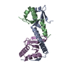
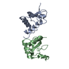
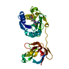
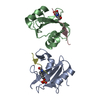
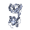
- PDB-3cpq: Crystal Structure of L30e a ribosomal protein from Methanocaldoco... -
+ Open data

- Basic information
| Entry | Database: PDB / ID: 3cpq | ||||||
|---|---|---|---|---|---|---|---|
| Title | Crystal Structure of L30e a ribosomal protein from Methanocaldococcus jannaschii DSM2661 (MJ1044) | ||||||
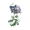
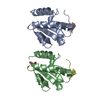
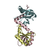
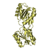
 Components Components | 50S ribosomal protein L30e Ribosome Ribosome | ||||||
 Keywords Keywords | RIBOSOMAL PROTEIN / ribosomal / RNA-protein / elongation factor / Ribonucleoprotein / Structural Genomics / NPPSFA / National Project on Protein Structural and Functional Analyses / RIKEN Structural Genomics/Proteomics Initiative / RSGI | ||||||
| Function / homology |  Function and homology information Function and homology informationcytosolic large ribosomal subunit / structural constituent of ribosome / translation / RNA bindingSimilarity search - Function | ||||||
| Biological species |   Methanocaldococcus jannaschii (archaea) Methanocaldococcus jannaschii (archaea) | ||||||
| Method | X-RAY DIFFRACTION / SYNCHROTRON / MOLECULAR REPLACEMENT / Resolution: 1.9 Å | ||||||
 Authors Authors | Jeyakanthan, J. / Sarani, R. / Mridula, P. / Sekar, K. / Kuramitsu, S. / Yokoyama, S. / RIKEN Structural Genomics/Proteomics Initiative (RSGI) | ||||||
 Citation Citation | Journal: To be Published Title: Crystal Structure of L30e a ribosomal protein from Methanocaldococcus jannaschii DSM2661 (MJ1044) Authors: Jeyakanthan, J. / Sarani, R. / Mridula, P. / Sekar, K. / Kuramitsu, S. / Yokoyama, S. | ||||||
| History |
|
- Structure visualization
| Structure viewer | Molecule: MolmilJmol/JSmol |
|---|
- Downloads & links
-Download
| PDBx/mmCIF format | 3cpq.cif.gz | 52.8 KB | Display | PDBx/mmCIF format |
|---|---|---|---|---|
| PDB format | pdb3cpq.ent.gz | 37.9 KB | Display | PDB format |
| PDBx/mmJSON format | 3cpq.json.gz | Tree view | PDBx/mmJSON format | |
| Others |  Other downloads Other downloads |
-Validation report
| Arichive directory | https://data.pdbj.org/pub/pdb/validation_reports/cp/3cpqftp://data.pdbj.org/pub/pdb/validation_reports/cp/3cpq | HTTPS FTP |
|---|
-Related structure data
| Related structure data | 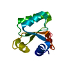 1w41S S: Starting model for refinement |
|---|---|
| Similar structure data |

-Links
 PDBj
PDBj


- Assembly
| Deposited unit | 
| ||||||||
|---|---|---|---|---|---|---|---|---|---|
| 1 |
| ||||||||
| 2 | 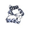
| ||||||||
| 3 | 
| ||||||||
| Unit cell |
|
-Components
| #1: Protein | Ribosome Mass: 12154.249 Da / Num. of mol.: 2 Source method: isolated from a genetically manipulated source Source: (gene. exp.) Methanocaldococcus jannaschii (archaea)Strain: DSM2661 / Plasmid: PET21A / Production host:  Escherichia coli (E. coli) / Strain (production host): ROSETTA(DE3) / References: UniProt: P54061 Escherichia coli (E. coli) / Strain (production host): ROSETTA(DE3) / References: UniProt: P54061#2: Water | ChemComp-HOH / | Water Mass: 18.015 Da / Num. of mol.: 127 / Source method: isolated from a natural source / Formula: H2O Mass: 18.015 Da / Num. of mol.: 127 / Source method: isolated from a natural source / Formula: H2O |
|---|
-Experimental details
-Experiment
| Experiment | Method: X-RAY DIFFRACTION / Number of used crystals: 1 |
|---|
- Sample preparation
| Crystal | Density Matthews: 2.15 Å3/Da / Density % sol: 42.91 % |
|---|---|
| Crystal grow | Temperature: 291 K / Method: microbatch / pH: 6 Details: 40% PEG400, 0.1 MES, 5% PEG3000, pH 6.0, MICROBATCH, temperature 291K |
-Data collection
| Diffraction | Mean temperature: 100 K |
|---|---|
| Diffraction source | Source: SYNCHROTRON / Site: SPring-8  / Beamline: BL26B1 / Wavelength: 1 Å / Beamline: BL26B1 / Wavelength: 1 Å |
| Detector | Type: RIGAKU / Detector: IMAGE PLATE / Date: Jun 13, 2007 / Details: RH Coated Bent-Cyrindrical MIRROR |
| Radiation | Monochromator: SI 1 1 1 DOUBLE CRYSTAL MONOCHROMATOR / Protocol: SINGLE WAVELENGTH / Monochromatic (M) / Laue (L): M / Scattering type: x-ray |
| Radiation wavelength | Wavelength: 1 Å / Relative weight: 1 |
| Reflection | Resolution: 1.9→50 Å / Num. obs: 16282 / % possible obs: 100 % / Biso Wilson estimate: 19.5 Å2 / Rmerge(I) obs: 0.067 / Rsym value: 0.088 |
| Reflection shell | Resolution: 1.9→1.97 Å / Rmerge(I) obs: 0.478 / Num. unique all: 1595 / Rsym value: 0.541 / % possible all: 100 |
- Processing
| Software |
| ||||||||||||||||||||
|---|---|---|---|---|---|---|---|---|---|---|---|---|---|---|---|---|---|---|---|---|---|
| Refinement | Method to determine structure: MOLECULAR REPLACEMENT Starting model: PDB ENTRY 1W41 Resolution: 1.9→46.12 Å / Rfactor Rfree error: 0.008 / Data cutoff high absF: 984491.78 / Data cutoff low absF: 0 / Isotropic thermal model: RESTRAINED / Cross valid method: THROUGHOUT / σ(F): 0 / Stereochemistry target values: Engh & Huber
| ||||||||||||||||||||
| Solvent computation | Solvent model: FLAT MODEL / Bsol: 64.0661 Å2 / ksol: 0.37649 e/Å3 | ||||||||||||||||||||
| Displacement parameters | Biso mean: 26.8 Å2
| ||||||||||||||||||||
| Refine analyze |
| ||||||||||||||||||||
| Refinement step | Cycle: LAST / Resolution: 1.9→46.12 Å
| ||||||||||||||||||||
| Refine LS restraints |
| ||||||||||||||||||||
| LS refinement shell | Resolution: 1.9→1.99 Å / Rfactor Rfree error: 0.027 / Total num. of bins used: 8
| ||||||||||||||||||||
| Xplor file |
|
