 Movie
Movie Controller


+ Open data

- Basic information
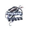
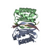
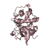
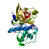
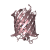
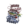
| Entry | Database: PDB / ID: 5e0l | ||||||
|---|---|---|---|---|---|---|---|
| Title | LC8 - Chica (415-424) Complex | ||||||
 Components Components |
| ||||||
 Keywords Keywords |  TRANSPORT PROTEIN / Complex / Dynein TRANSPORT PROTEIN / Complex / Dynein | ||||||
| Function / homology |  Function and homology information Function and homology informationprotein localization to mitotic spindle / spermatid nucleus elongation / chaeta morphogenesis / positive regulation of neuron remodeling / Macroautophagy / Aggrephagy / wing disc development / HSP90 chaperone cycle for steroid hormone receptors (SHR) in the presence of ligand / COPI-mediated anterograde transport / COPI-independent Golgi-to-ER retrograde traffic ...protein localization to mitotic spindle / spermatid nucleus elongation / chaeta morphogenesis / positive regulation of neuron remodeling / Macroautophagy / Aggrephagy / wing disc development / HSP90 chaperone cycle for steroid hormone receptors (SHR) in the presence of ligand / COPI-mediated anterograde transport / COPI-independent Golgi-to-ER retrograde traffic / : / chaeta development / sperm individualization / microtubule anchoring at centrosome / metaphase chromosome alignment / imaginal disc-derived wing morphogenesis / positive regulation of cell cycle G1/S phase transition / Neutrophil degranulation / regulation of TOR signaling / dynein complex / cytoplasmic dynein complex / dynein light intermediate chain binding / dynein intermediate chain binding / oogenesis / intercellular bridge / mitotic spindle pole / regulation of protein catabolic process / establishment of mitotic spindle orientation / actin filament bundle assembly / kinesin binding / epithelial to mesenchymal transition / centriole / regulation of ERK1 and ERK2 cascade / mitotic spindle / spindle / autophagy / microtubule cytoskeleton / disordered domain specific binding / cell migration / microtubule binding / spermatogenesis / cell population proliferation / microtubule / cell division / protein kinase binding / signal transduction / protein homodimerization activity / protein-containing complex / cytosol / cytoplasmSimilarity search - Function | ||||||
| Biological species |  Drosophila melanogaster (fruit fly) Drosophila melanogaster (fruit fly) | ||||||
| Method | X-RAY DIFFRACTION / SYNCHROTRON / MOLECULAR REPLACEMENT / Resolution: 1.31 Å | ||||||
 Authors Authors | Clark, S.A. / Barbar, E.B. / Karplus, P.A. | ||||||
 Citation Citation | Journal: Biochemistry / Year: 2016 Title: The Anchored Flexibility Model in LC8 Motif Recognition: Insights from the Chica Complex. Authors: Clark, S. / Nyarko, A. / Lohr, F. / Karplus, P.A. / Barbar, E. | ||||||
| History |
|
- Structure visualization
| Structure viewer | Molecule: MolmilJmol/JSmol |
|---|
- Downloads & links
-Download
| PDBx/mmCIF format | 5e0l.cif.gz | 75.8 KB | Display | PDBx/mmCIF format |
|---|---|---|---|---|
| PDB format | pdb5e0l.ent.gz | 56.6 KB | Display | PDB format |
| PDBx/mmJSON format | 5e0l.json.gz | Tree view | PDBx/mmJSON format | |
| Others |  Other downloads Other downloads |
-Validation report
| Arichive directory | https://data.pdbj.org/pub/pdb/validation_reports/e0/5e0lftp://data.pdbj.org/pub/pdb/validation_reports/e0/5e0l | HTTPS FTP |
|---|
-Related structure data
| Related structure data |  5e0mC  3briS C: citing same article ( S: Starting model for refinement |
|---|---|
| Similar structure data |


-Links
 PDBj
PDBj


- Assembly
| Deposited unit | 
| ||||||||
|---|---|---|---|---|---|---|---|---|---|
| 1 | 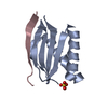
| ||||||||
| Unit cell |
| ||||||||
| Components on special symmetry positions |
|
-Components
| #1: Protein | Mass: 10388.849 Da / Num. of mol.: 1 Source method: isolated from a genetically manipulated source Source: (gene. exp.) Drosophila melanogaster (fruit fly) / Gene: ctp, Cdlc1, ddlc1, CG6998 / Production host:  Escherichia coli (E. coli) / References: UniProt: Q24117 Escherichia coli (E. coli) / References: UniProt: Q24117 |
|---|---|
| #2: Protein/peptide | Mass: 1584.684 Da / Num. of mol.: 1 Source method: isolated from a genetically manipulated source Source: (gene. exp.) Drosophila melanogaster (fruit fly) / Production host: Escherichia coli (E. coli) / References: UniProt: Q9H4H8*PLUS |
| #3: Chemical | ChemComp-SO4 / Sulfate  Mass: 96.063 Da / Num. of mol.: 1 / Source method: obtained synthetically / Formula: SO4 Mass: 96.063 Da / Num. of mol.: 1 / Source method: obtained synthetically / Formula: SO4 |
| #4: Water | ChemComp-HOH / Water Mass: 18.015 Da / Num. of mol.: 59 / Source method: isolated from a natural source / Formula: H2O Mass: 18.015 Da / Num. of mol.: 59 / Source method: isolated from a natural source / Formula: H2O |
-Experimental details
-Experiment
| Experiment | Method: X-RAY DIFFRACTION |
|---|
- Sample preparation
| Crystal | Density Matthews: 2.59 Å3/Da / Density % sol: 52.56 % / Description: Long hexagonal rods |
|---|---|
| Crystal grow | Temperature: 298 K / Method: vapor diffusion, hanging drop / pH: 5.5 Details: 0.1 M calcium acetate, 0.1 M sodium cacodylate, 15% polyethylene glycol 8000 |
-Data collection
| Diffraction | Mean temperature: 100 K |
|---|---|
| Diffraction source | Source: SYNCHROTRON / Site: ALS  / Beamline: 5.0.2 / Wavelength: 1 Å / Beamline: 5.0.2 / Wavelength: 1 Å |
| Detector | Type: ADSC QUANTUM 315r / Detector: CCD / Date: Feb 8, 2015 |
| Radiation | Protocol: SINGLE WAVELENGTH / Monochromatic (M) / Laue (L): M / Scattering type: x-ray |
| Radiation wavelength | Wavelength: 1 Å / Relative weight: 1 |
| Reflection | Resolution: 1.31→38.7 Å / Num. obs: 30438 / % possible obs: 100 % / Redundancy: 30.8 % / Rmerge(I) obs: 0.081 / Net I/σ(I): 20.6 |
| Reflection shell | Resolution: 1.31→1.33 Å / Redundancy: 8 % / Rmerge(I) obs: 4.45 / Mean I/σ(I) obs: 0.4 / % possible all: 99.1 |
- Processing
| Software |
| ||||||||||||||||||||||||||||||||||||||||||||||||||||||||||||||||||||||||||||||||||||
|---|---|---|---|---|---|---|---|---|---|---|---|---|---|---|---|---|---|---|---|---|---|---|---|---|---|---|---|---|---|---|---|---|---|---|---|---|---|---|---|---|---|---|---|---|---|---|---|---|---|---|---|---|---|---|---|---|---|---|---|---|---|---|---|---|---|---|---|---|---|---|---|---|---|---|---|---|---|---|---|---|---|---|---|---|---|
| Refinement | Method to determine structure: MOLECULAR REPLACEMENT Starting model: 3BRI Resolution: 1.31→38.7 Å / SU ML: 0.19 / Cross valid method: FREE R-VALUE / σ(F): 1.34 / Phase error: 28.59 / Stereochemistry target values: ML
| ||||||||||||||||||||||||||||||||||||||||||||||||||||||||||||||||||||||||||||||||||||
| Solvent computation | Shrinkage radii: 0.9 Å / VDW probe radii: 1.11 Å / Solvent model: FLAT BULK SOLVENT MODEL | ||||||||||||||||||||||||||||||||||||||||||||||||||||||||||||||||||||||||||||||||||||
| Refinement step | Cycle: LAST / Resolution: 1.31→38.7 Å
| ||||||||||||||||||||||||||||||||||||||||||||||||||||||||||||||||||||||||||||||||||||
| Refine LS restraints |
| ||||||||||||||||||||||||||||||||||||||||||||||||||||||||||||||||||||||||||||||||||||
| LS refinement shell |
| ||||||||||||||||||||||||||||||||||||||||||||||||||||||||||||||||||||||||||||||||||||
| Refinement TLS params. | Method: refined / Origin x: -17.0657 Å / Origin y: 15.4526 Å / Origin z: -8.4479 Å
| ||||||||||||||||||||||||||||||||||||||||||||||||||||||||||||||||||||||||||||||||||||
| Refinement TLS group | Selection details: all |
