 Movie
Movie Controller


+ Open data

- Basic information
| Entry | Database: PDB / ID: 7k09 | ||||||
|---|---|---|---|---|---|---|---|
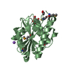
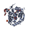
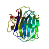
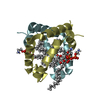
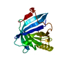
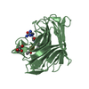
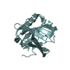
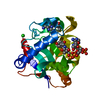
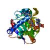
| Title | Puromycin N-acetyltransferase in complex with acetyl-CoA | ||||||
 Components Components | Puromycin N-acetyltransferase | ||||||
 Keywords Keywords |  TRANSFERASE / Enzyme acetyletransferase complex selection marker / ANTIBIOTIC TRANSFERASE / Enzyme acetyletransferase complex selection marker / ANTIBIOTIC | ||||||
| Function / homology | Transferases; Acyltransferases / Acetyltransferase (GNAT) family / acetyltransferase activity / Gcn5-related N-acetyltransferase (GNAT) domain profile. / GNAT domain / Acyl-CoA N-acyltransferase / response to antibiotic / ACETYL COENZYME *A / Puromycin N-acetyltransferase Function and homology information Function and homology information | ||||||
| Biological species |  Streptomyces alboniger (bacteria) Streptomyces alboniger (bacteria) | ||||||
| Method | X-RAY DIFFRACTION / SYNCHROTRON / MOLECULAR REPLACEMENT / Resolution: 2.313 Å | ||||||
 Authors Authors | Caputo, A.T. / Newman, J. / Adams, T.E. / Peat, T.S. | ||||||
 Citation Citation | Journal: Sci Rep / Year: 2021 Title: Structure-guided selection of puromycin N-acetyltransferase mutants with enhanced selection stringency for deriving mammalian cell lines expressing recombinant proteins. Authors: Caputo, A.T. / Eder, O.M. / Bereznakova, H. / Pothuis, H. / Ardevol, A. / Newman, J. / Nuttall, S. / Peat, T.S. / Adams, T.E. | ||||||
| History |
|
- Structure visualization
| Structure viewer | Molecule: MolmilJmol/JSmol |
|---|
- Downloads & links
-Download
| PDBx/mmCIF format | 7k09.cif.gz | 642 KB | Display | PDBx/mmCIF format |
|---|---|---|---|---|
| PDB format | pdb7k09.ent.gz | 551.7 KB | Display | PDB format |
| PDBx/mmJSON format | 7k09.json.gz | Tree view | PDBx/mmJSON format | |
| Others |  Other downloads Other downloads |
-Validation report
| Arichive directory | https://data.pdbj.org/pub/pdb/validation_reports/k0/7k09ftp://data.pdbj.org/pub/pdb/validation_reports/k0/7k09 | HTTPS FTP |
|---|
-Related structure data
| Related structure data |  7k0aC 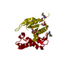 2qecS S: Starting model for refinement C: citing same article ( |
|---|---|
| Similar structure data |

-Links
 PDBj
PDBj

- Assembly
| Deposited unit | 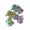
| ||||||||
|---|---|---|---|---|---|---|---|---|---|
| 1 | 
| ||||||||
| 2 | 
| ||||||||
| 3 | 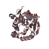
| ||||||||
| 4 | 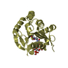
| ||||||||
| 5 | 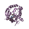
| ||||||||
| 6 | 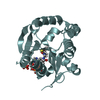
| ||||||||
| Unit cell |
|
-Components
| #1: Protein | Mass: 22581.490 Da / Num. of mol.: 6 Source method: isolated from a genetically manipulated source Source: (gene. exp.) Streptomyces alboniger (bacteria) / Gene: pac / Plasmid: pET43 / Production host: Escherichia coli BL21(DE3) (bacteria) / References: UniProt: P13249, Transferases; Acyltransferases#2: Chemical | ChemComp-ACO / Acetyl-CoA  Mass: 809.571 Da / Num. of mol.: 6 / Source method: obtained synthetically / Formula: C23H38N7O17P3S / Feature type: SUBJECT OF INVESTIGATION Mass: 809.571 Da / Num. of mol.: 6 / Source method: obtained synthetically / Formula: C23H38N7O17P3S / Feature type: SUBJECT OF INVESTIGATION#3: Water | ChemComp-HOH / | Water Mass: 18.015 Da / Num. of mol.: 24 / Source method: isolated from a natural source / Formula: H2O Mass: 18.015 Da / Num. of mol.: 24 / Source method: isolated from a natural source / Formula: H2OHas ligand of interest | Y | |
|---|
-Experimental details
-Experiment
| Experiment | Method: X-RAY DIFFRACTION / Number of used crystals: 1 |
|---|
- Sample preparation
| Crystal | Density Matthews: 2.15 Å3/Da / Density % sol: 42.84 % / Description: small 20 um chunks |
|---|---|
| Crystal grow | Temperature: 281 K / Method: vapor diffusion, sitting drop / pH: 9 Details: 2.5 M ammonium sulfate, 10% v/v DL-malate-MES-Tris pH 9.0 |
-Data collection
| Diffraction | Mean temperature: 100 K / Serial crystal experiment: N |
|---|---|
| Diffraction source | Source: SYNCHROTRON / Site: Australian Synchrotron  / Beamline: MX2 / Wavelength: 0.95372 Å / Beamline: MX2 / Wavelength: 0.95372 Å |
| Detector | Type: DECTRIS EIGER X 16M / Detector: PIXEL / Date: Jun 19, 2019 |
| Radiation | Protocol: SINGLE WAVELENGTH / Monochromatic (M) / Laue (L): M / Scattering type: x-ray |
| Radiation wavelength | Wavelength: 0.95372 Å / Relative weight: 1 |
| Reflection | Resolution: 2.361→106.242 Å / Num. obs: 27792 / % possible obs: 93.1 % / Redundancy: 16.9 % / CC1/2: 0.994 / Rmerge(I) obs: 0.502 / Rpim(I) all: 0.125 / Rrim(I) all: 0.517 / Net I/σ(I): 7.7 |
| Reflection shell | Resolution: 2.361→2.589 Å / Redundancy: 15.8 % / Mean I/σ(I) obs: 1.5 / Num. unique obs: 1192 / CC1/2: 0.504 / Rpim(I) all: 0.795 / % possible all: 67 |
- Processing
| Software |
| |||||||||||||||||||||||||||||||||||||||||||||||||||||||||||||||||||||||||||||||||||||||||||||||||||||||||||||||||||||||||||||||||||||||||||||||||||||||||||||||||||||||||||||||
|---|---|---|---|---|---|---|---|---|---|---|---|---|---|---|---|---|---|---|---|---|---|---|---|---|---|---|---|---|---|---|---|---|---|---|---|---|---|---|---|---|---|---|---|---|---|---|---|---|---|---|---|---|---|---|---|---|---|---|---|---|---|---|---|---|---|---|---|---|---|---|---|---|---|---|---|---|---|---|---|---|---|---|---|---|---|---|---|---|---|---|---|---|---|---|---|---|---|---|---|---|---|---|---|---|---|---|---|---|---|---|---|---|---|---|---|---|---|---|---|---|---|---|---|---|---|---|---|---|---|---|---|---|---|---|---|---|---|---|---|---|---|---|---|---|---|---|---|---|---|---|---|---|---|---|---|---|---|---|---|---|---|---|---|---|---|---|---|---|---|---|---|---|---|---|---|---|
| Refinement | Method to determine structure: MOLECULAR REPLACEMENT Starting model: 2QEC Resolution: 2.313→106.24 Å / Cor.coef. Fo:Fc: 0.882 / Cor.coef. Fo:Fc free: 0.867 / Cross valid method: THROUGHOUT / SU Rfree Blow DPI: 0.4
| |||||||||||||||||||||||||||||||||||||||||||||||||||||||||||||||||||||||||||||||||||||||||||||||||||||||||||||||||||||||||||||||||||||||||||||||||||||||||||||||||||||||||||||||
| Displacement parameters | Biso mean: 45.44 Å2
| |||||||||||||||||||||||||||||||||||||||||||||||||||||||||||||||||||||||||||||||||||||||||||||||||||||||||||||||||||||||||||||||||||||||||||||||||||||||||||||||||||||||||||||||
| Refine analyze | Luzzati coordinate error obs: 0.46 Å | |||||||||||||||||||||||||||||||||||||||||||||||||||||||||||||||||||||||||||||||||||||||||||||||||||||||||||||||||||||||||||||||||||||||||||||||||||||||||||||||||||||||||||||||
| Refinement step | Cycle: LAST / Resolution: 2.313→106.24 Å
| |||||||||||||||||||||||||||||||||||||||||||||||||||||||||||||||||||||||||||||||||||||||||||||||||||||||||||||||||||||||||||||||||||||||||||||||||||||||||||||||||||||||||||||||
| Refine LS restraints |
| |||||||||||||||||||||||||||||||||||||||||||||||||||||||||||||||||||||||||||||||||||||||||||||||||||||||||||||||||||||||||||||||||||||||||||||||||||||||||||||||||||||||||||||||
| LS refinement shell | Resolution: 2.361→2.5 Å
| |||||||||||||||||||||||||||||||||||||||||||||||||||||||||||||||||||||||||||||||||||||||||||||||||||||||||||||||||||||||||||||||||||||||||||||||||||||||||||||||||||||||||||||||
| Refinement TLS params. | Refine-ID: X-RAY DIFFRACTION
| |||||||||||||||||||||||||||||||||||||||||||||||||||||||||||||||||||||||||||||||||||||||||||||||||||||||||||||||||||||||||||||||||||||||||||||||||||||||||||||||||||||||||||||||
| Refinement TLS group |
|
