 Movie
Movie Controller

[English] 日本語
 Yorodumi
- PDB-2ifw: Crystal structure of scytalido-glutamic peptidase with a transiti... -
+ Open data

- Basic information
| Entry | Database: PDB / ID: 2ifw | ||||||
|---|---|---|---|---|---|---|---|
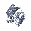
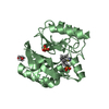
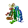
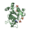
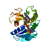
| Title | Crystal structure of scytalido-glutamic peptidase with a transition state analog inhibitor | ||||||
 Components Components |
| ||||||
 Keywords Keywords | HYDROLASE/HYDROLASE INHIBITOR / enzyme-inhibitor complex / HYDROLASE-HYDROLASE INHIBITOR complex | ||||||
| Function / homology |  Function and homology information Function and homology information scytalidopepsin B / glutamic-type endopeptidase activity / aspartic-type endopeptidase activity / proteolysis scytalidopepsin B / glutamic-type endopeptidase activity / aspartic-type endopeptidase activity / proteolysisSimilarity search - Function | ||||||
| Biological species |  Scytalidium lignicola (fungus) Scytalidium lignicola (fungus) | ||||||
| Method | X-RAY DIFFRACTION / SYNCHROTRON / MOLECULAR REPLACEMENT / Resolution: 2.3 Å | ||||||
 Authors Authors | Pillai, B. / Cherney, M.M. / Hiraga, K. / Takada, K. / Oda, K. / James, M.N. | ||||||
 Citation Citation | Journal: J.Mol.Biol. / Year: 2007 Title: Crystal structure of scytalidoglutamic peptidase with its first potent inhibitor provides insights into substrate specificity and catalysis. Authors: Pillai, B. / Cherney, M.M. / Hiraga, K. / Takada, K. / Oda, K. / James, M.N. | ||||||
| History |
|
- Structure visualization
| Structure viewer | Molecule: MolmilJmol/JSmol |
|---|
- Downloads & links
-Download
| PDBx/mmCIF format | 2ifw.cif.gz | 94.2 KB | Display | PDBx/mmCIF format |
|---|---|---|---|---|
| PDB format | pdb2ifw.ent.gz | 71.9 KB | Display | PDB format |
| PDBx/mmJSON format | 2ifw.json.gz | Tree view | PDBx/mmJSON format | |
| Others |  Other downloads Other downloads |
-Validation report
| Arichive directory | https://data.pdbj.org/pub/pdb/validation_reports/if/2ifwftp://data.pdbj.org/pub/pdb/validation_reports/if/2ifw | HTTPS FTP |
|---|
-Related structure data
| Related structure data | 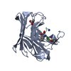 2ifrC 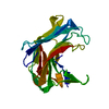 1s2bS S: Starting model for refinement C: citing same article ( |
|---|---|
| Similar structure data |


-Links
 PDBj
PDBj

- Assembly
| Deposited unit | 
| ||||||||
|---|---|---|---|---|---|---|---|---|---|
| 1 | 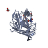
| ||||||||
| 2 | 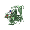
| ||||||||
| Unit cell |
|
-Components
| #1: Protein | / Acid protease B / SLB Mass: 21553.812 Da / Num. of mol.: 2 Source method: isolated from a genetically manipulated source Source: (gene. exp.) Scytalidium lignicola (fungus) / Production host:  Escherichia coli (E. coli) / References: UniProt: P15369, scytalidopepsin B Escherichia coli (E. coli) / References: UniProt: P15369, scytalidopepsin B#2: Protein/peptide | PeptideMass: 928.174 Da / Num. of mol.: 2 / Source method: obtained synthetically #3: Chemical | Acetic acid  Mass: 60.052 Da / Num. of mol.: 2 / Source method: obtained synthetically / Formula: C2H4O2 Mass: 60.052 Da / Num. of mol.: 2 / Source method: obtained synthetically / Formula: C2H4O2#4: Chemical | Glycerol  Mass: 92.094 Da / Num. of mol.: 2 / Source method: obtained synthetically / Formula: C3H8O3 Mass: 92.094 Da / Num. of mol.: 2 / Source method: obtained synthetically / Formula: C3H8O3#5: Water | ChemComp-HOH / | Water Mass: 18.015 Da / Num. of mol.: 111 / Source method: isolated from a natural source / Formula: H2O Mass: 18.015 Da / Num. of mol.: 111 / Source method: isolated from a natural source / Formula: H2O |
|---|
-Experimental details
-Experiment
| Experiment | Method: X-RAY DIFFRACTION / Number of used crystals: 1 |
|---|
- Sample preparation
| Crystal | Density Matthews: 2.17 Å3/Da / Density % sol: 43.22 % |
|---|---|
| Crystal grow | Temperature: 298 K / Method: vapor diffusion, sitting drop / pH: 5.5 Details: 25% PEG 3300, 0.2 M NaCl, 0.1 M Bis-Tris buffer, pH 5.5, VAPOR DIFFUSION, SITTING DROP, temperature 298K |
-Data collection
| Diffraction | Mean temperature: 100 K |
|---|---|
| Diffraction source | Source: SYNCHROTRON / Site: ALS  / Beamline: 8.3.1 / Wavelength: 1.115869 Å / Beamline: 8.3.1 / Wavelength: 1.115869 Å |
| Detector | Type: ADSC QUANTUM 210 / Detector: CCD / Date: Apr 20, 2005 |
| Radiation | Monochromator: double crystal / Protocol: SINGLE WAVELENGTH / Monochromatic (M) / Laue (L): M / Scattering type: x-ray |
| Radiation wavelength | Wavelength: 1.115869 Å / Relative weight: 1 |
| Reflection | Resolution: 2.3→30 Å / Num. all: 17716 / Num. obs: 17716 / % possible obs: 98.1 % / Observed criterion σ(F): 0 / Observed criterion σ(I): 0 |
| Reflection shell | Resolution: 2.3→2.38 Å / % possible all: 96.5 |
- Processing
| Software |
| |||||||||||||||||||||||||
|---|---|---|---|---|---|---|---|---|---|---|---|---|---|---|---|---|---|---|---|---|---|---|---|---|---|---|
| Refinement | Method to determine structure: MOLECULAR REPLACEMENT Starting model: pdb id 1S2B Resolution: 2.3→30 Å / σ(F): 0 / σ(I): 0 / Stereochemistry target values: Engh & Huber
| |||||||||||||||||||||||||
| Refinement step | Cycle: LAST / Resolution: 2.3→30 Å
| |||||||||||||||||||||||||
| Refine LS restraints |
|
