 Movie
Movie Controller


+ Open data

- Basic information
| Entry | Database: PDB / ID: 5h5s | ||||||
|---|---|---|---|---|---|---|---|
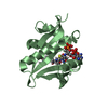
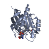
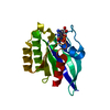
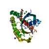
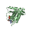
| Title | Crystal structure of human GPX4 in complex with GXpep-3 | ||||||
 Components Components |
| ||||||
 Keywords Keywords | OXIDOREDUCTASE/OXIDOREDUCTASE INHIBITOR / PHOSPHOLIPID HYDROPEROXIDE GLUTATHIONE PEROXIDASE 4 /  SELENOCYSTEINE / OXIDOREDUCTASE / OXIDOREDUCTASE-OXIDOREDUCTASE INHIBITOR complex SELENOCYSTEINE / OXIDOREDUCTASE / OXIDOREDUCTASE-OXIDOREDUCTASE INHIBITOR complex | ||||||
| Function / homology |  Function and homology informationphospholipid-hydroperoxide glutathione peroxidase / phospholipid-hydroperoxide glutathione peroxidase activity / Synthesis of 15-eicosatetraenoic acid derivatives / Synthesis of 12-eicosatetraenoic acid derivatives / negative regulation of ferroptosis / selenium binding / glutathione peroxidase / Synthesis of 5-eicosatetraenoic acids / lipoxygenase pathway / arachidonic acid metabolic process ...phospholipid-hydroperoxide glutathione peroxidase / phospholipid-hydroperoxide glutathione peroxidase activity / Synthesis of 15-eicosatetraenoic acid derivatives / Synthesis of 12-eicosatetraenoic acid derivatives / negative regulation of ferroptosis / selenium binding / glutathione peroxidase / Synthesis of 5-eicosatetraenoic acids / lipoxygenase pathway / arachidonic acid metabolic process / Biosynthesis of aspirin-triggered D-series resolvins / Biosynthesis of E-series 18(R)-resolvins / Biosynthesis of D-series resolvins / Biosynthesis of E-series 18(S)-resolvins / glutathione peroxidase activity / long-chain fatty acid biosynthetic process / protein polymerization / phospholipid metabolic process / response to estradiol / nuclear envelope / chromatin organization / cellular response to oxidative stress / spermatogenesis / response to oxidative stress / protein-containing complex / mitochondrion / extracellular exosome / identical protein binding / nucleus / cytosol Function and homology informationphospholipid-hydroperoxide glutathione peroxidase / phospholipid-hydroperoxide glutathione peroxidase activity / Synthesis of 15-eicosatetraenoic acid derivatives / Synthesis of 12-eicosatetraenoic acid derivatives / negative regulation of ferroptosis / selenium binding / glutathione peroxidase / Synthesis of 5-eicosatetraenoic acids / lipoxygenase pathway / arachidonic acid metabolic process ...phospholipid-hydroperoxide glutathione peroxidase / phospholipid-hydroperoxide glutathione peroxidase activity / Synthesis of 15-eicosatetraenoic acid derivatives / Synthesis of 12-eicosatetraenoic acid derivatives / negative regulation of ferroptosis / selenium binding / glutathione peroxidase / Synthesis of 5-eicosatetraenoic acids / lipoxygenase pathway / arachidonic acid metabolic process / Biosynthesis of aspirin-triggered D-series resolvins / Biosynthesis of E-series 18(R)-resolvins / Biosynthesis of D-series resolvins / Biosynthesis of E-series 18(S)-resolvins / glutathione peroxidase activity / long-chain fatty acid biosynthetic process / protein polymerization / phospholipid metabolic process / response to estradiol / nuclear envelope / chromatin organization / cellular response to oxidative stress / spermatogenesis / response to oxidative stress / protein-containing complex / mitochondrion / extracellular exosome / identical protein binding / nucleus / cytosolSimilarity search - Function | ||||||
| Biological species |  Homo sapiens (human) Homo sapiens (human)  Enterobacteria phage T7 (virus) Enterobacteria phage T7 (virus) | ||||||
| Method | X-RAY DIFFRACTION / SYNCHROTRON / MOLECULAR REPLACEMENT / Resolution: 1.85 Å | ||||||
 Authors Authors | Sogabe, S. / Kadotani, A. / Lane, W. / Snell, G. | ||||||
 Citation Citation | Journal: Biochem. Biophys. Res. Commun. / Year: 2017 Title: Discovery of GPX4 inhibitory peptides from random peptide T7 phage display and subsequent structural analysis Authors: Sakamoto, K. / Sogabe, S. / Kamada, Y. / Matsumoto, S.I. / Kadotani, A. / Sakamoto, J.I. / Tani, A. | ||||||
| History |
|
- Structure visualization
| Structure viewer | Molecule: MolmilJmol/JSmol |
|---|
- Downloads & links
-Download
| PDBx/mmCIF format | 5h5s.cif.gz | 89.1 KB | Display | PDBx/mmCIF format |
|---|---|---|---|---|
| PDB format | pdb5h5s.ent.gz | 65.1 KB | Display | PDB format |
| PDBx/mmJSON format | 5h5s.json.gz | Tree view | PDBx/mmJSON format | |
| Others |  Other downloads Other downloads |
-Validation report
| Arichive directory | https://data.pdbj.org/pub/pdb/validation_reports/h5/5h5sftp://data.pdbj.org/pub/pdb/validation_reports/h5/5h5s | HTTPS FTP |
|---|
-Related structure data
| Related structure data |  5h5qC  5h5rC 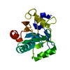 2obiS C: citing same article ( S: Starting model for refinement |
|---|---|
| Similar structure data |


-Links
 PDBj
PDBj
- Assembly
| Deposited unit | 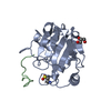
| ||||||||
|---|---|---|---|---|---|---|---|---|---|
| 1 |
| ||||||||
| Unit cell |
|
-Components
| #1: Protein | Mass: 19403.070 Da / Num. of mol.: 1 / Mutation: C29S, C37A, C64S, U73C, C93R, C102S, C134E, C175V Source method: isolated from a genetically manipulated source Source: (gene. exp.) Homo sapiens (human) / Gene: GPX4 / Production host:  Escherichia coli (E. coli) Escherichia coli (E. coli)References: UniProt: P36969, phospholipid-hydroperoxide glutathione peroxidase |
|---|---|
| #2: Protein/peptide | Mass: 1561.887 Da / Num. of mol.: 1 / Source method: obtained synthetically / Source: (synth.) Enterobacteria phage T7 (virus) |
| #3: Chemical | ChemComp-GOL / Glycerol  Mass: 92.094 Da / Num. of mol.: 1 / Source method: obtained synthetically / Formula: C3H8O3 Mass: 92.094 Da / Num. of mol.: 1 / Source method: obtained synthetically / Formula: C3H8O3 |
| #4: Water | ChemComp-HOH / Water Mass: 18.015 Da / Num. of mol.: 126 / Source method: isolated from a natural source / Formula: H2O Mass: 18.015 Da / Num. of mol.: 126 / Source method: isolated from a natural source / Formula: H2O |
-Experimental details
-Experiment
| Experiment | Method: X-RAY DIFFRACTION / Number of used crystals: 1 |
|---|
- Sample preparation
| Crystal | Density Matthews: 1.92 Å3/Da / Density % sol: 35.94 % |
|---|---|
| Crystal grow | Temperature: 293 K / Method: vapor diffusion, sitting drop / pH: 5.5 / Details: 0.1M MES pH 5.5, 25% PEG 4000 |
-Data collection
| Diffraction | Mean temperature: 100 K |
|---|---|
| Diffraction source | Source: SYNCHROTRON / Site: Photon Factory  / Beamline: BL-17A / Wavelength: 1 Å / Beamline: BL-17A / Wavelength: 1 Å |
| Detector | Type: DECTRIS PILATUS3 6M / Detector: PIXEL / Date: Jun 15, 2016 |
| Radiation | Protocol: SINGLE WAVELENGTH / Monochromatic (M) / Laue (L): M / Scattering type: x-ray |
| Radiation wavelength | Wavelength: 1 Å / Relative weight: 1 |
| Reflection | Resolution: 1.85→50 Å / Num. obs: 13498 / % possible obs: 97.8 % / Redundancy: 3.1 % / Rmerge(I) obs: 0.098 / Net I/σ(I): 12.1 |
| Reflection shell | Resolution: 1.85→1.88 Å / Redundancy: 1.9 % / Rmerge(I) obs: 0.386 / Mean I/σ(I) obs: 1.8 / % possible all: 76.7 |
- Processing
| Software |
| ||||||||||||||||||||||||||||||||||||||||||||||||||||||||||||||||||||||||||||||||||||||||||||||||||||||||||||||||||||||||||||||||||||||||||||||||||||||||||||||||||||||||||||||||||||||
|---|---|---|---|---|---|---|---|---|---|---|---|---|---|---|---|---|---|---|---|---|---|---|---|---|---|---|---|---|---|---|---|---|---|---|---|---|---|---|---|---|---|---|---|---|---|---|---|---|---|---|---|---|---|---|---|---|---|---|---|---|---|---|---|---|---|---|---|---|---|---|---|---|---|---|---|---|---|---|---|---|---|---|---|---|---|---|---|---|---|---|---|---|---|---|---|---|---|---|---|---|---|---|---|---|---|---|---|---|---|---|---|---|---|---|---|---|---|---|---|---|---|---|---|---|---|---|---|---|---|---|---|---|---|---|---|---|---|---|---|---|---|---|---|---|---|---|---|---|---|---|---|---|---|---|---|---|---|---|---|---|---|---|---|---|---|---|---|---|---|---|---|---|---|---|---|---|---|---|---|---|---|---|---|
| Refinement | Method to determine structure: MOLECULAR REPLACEMENT Starting model: 2OBI Resolution: 1.85→40 Å / Cor.coef. Fo:Fc: 0.958 / Cor.coef. Fo:Fc free: 0.941 / SU B: 6.569 / SU ML: 0.099 / Cross valid method: THROUGHOUT / ESU R: 0.155 / ESU R Free: 0.136 / Details: HYDROGENS HAVE BEEN ADDED IN THE RIDING POSITIONS
| ||||||||||||||||||||||||||||||||||||||||||||||||||||||||||||||||||||||||||||||||||||||||||||||||||||||||||||||||||||||||||||||||||||||||||||||||||||||||||||||||||||||||||||||||||||||
| Solvent computation | Ion probe radii: 0.8 Å / Shrinkage radii: 0.8 Å / VDW probe radii: 1.2 Å | ||||||||||||||||||||||||||||||||||||||||||||||||||||||||||||||||||||||||||||||||||||||||||||||||||||||||||||||||||||||||||||||||||||||||||||||||||||||||||||||||||||||||||||||||||||||
| Displacement parameters | Biso mean: 16.791 Å2
| ||||||||||||||||||||||||||||||||||||||||||||||||||||||||||||||||||||||||||||||||||||||||||||||||||||||||||||||||||||||||||||||||||||||||||||||||||||||||||||||||||||||||||||||||||||||
| Refinement step | Cycle: 1 / Resolution: 1.85→40 Å
| ||||||||||||||||||||||||||||||||||||||||||||||||||||||||||||||||||||||||||||||||||||||||||||||||||||||||||||||||||||||||||||||||||||||||||||||||||||||||||||||||||||||||||||||||||||||
| Refine LS restraints |
|