 Movie
Movie Controller

[English] 日本語
 Yorodumi
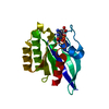
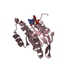
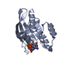
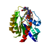
- PDB-2fe4: The crystal structure of human neuronal Rab6B in its inactive GDP... -
+ Open data

- Basic information
| Entry | Database: PDB / ID: 2fe4 | ||||||
|---|---|---|---|---|---|---|---|
| Title | The crystal structure of human neuronal Rab6B in its inactive GDP-bound form | ||||||
 Components Components | Ras-related protein Rab-6B | ||||||
 Keywords Keywords |  HYDROLASE / protein-nucleotide complex HYDROLASE / protein-nucleotide complex | ||||||
| Function / homology |  Function and homology information Function and homology informationprotein localization to Golgi membrane / Retrograde transport at the Trans-Golgi-Network / intra-Golgi vesicle-mediated transport / RAB geranylgeranylation / myosin V binding / COPI-independent Golgi-to-ER retrograde traffic / RAB GEFs exchange GTP for GDP on RABs / retrograde vesicle-mediated transport, Golgi to endoplasmic reticulum / retrograde transport, endosome to Golgi / TBC/RABGAPs ...protein localization to Golgi membrane / Retrograde transport at the Trans-Golgi-Network / intra-Golgi vesicle-mediated transport / RAB geranylgeranylation / myosin V binding / COPI-independent Golgi-to-ER retrograde traffic / RAB GEFs exchange GTP for GDP on RABs / retrograde vesicle-mediated transport, Golgi to endoplasmic reticulum / retrograde transport, endosome to Golgi / TBC/RABGAPs / Golgi organization / endoplasmic reticulum-Golgi intermediate compartment / endomembrane system / small monomeric GTPase / intracellular protein transport / neuron projection development / presynapse / cytoplasmic vesicle / Golgi membrane / GTPase activity / GTP binding / Golgi apparatus / cytosolSimilarity search - Function | ||||||
| Biological species |  Homo sapiens (human) Homo sapiens (human) | ||||||
| Method | X-RAY DIFFRACTION / SYNCHROTRON / Rab6 from Plasmodium falciparum (PDB code 1D5C) / Resolution: 2.3 Å | ||||||
 Authors Authors | Garcia-Saez, I. / Tcherniuk, F. / Kozielski, F. | ||||||
 Citation Citation | Journal: Acta Crystallogr.,Sect.D / Year: 2006 Title: The structure of human neuronal Rab6B in the active and inactive form. Authors: Garcia-Saez, I. / Tcherniuk, S. / Kozielski, F. | ||||||
| History |
|
- Structure visualization
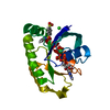
| Structure viewer | Molecule: MolmilJmol/JSmol |
|---|
- Downloads & links
-Download
| PDBx/mmCIF format | 2fe4.cif.gz | 50 KB | Display | PDBx/mmCIF format |
|---|---|---|---|---|
| PDB format | pdb2fe4.ent.gz | 34.8 KB | Display | PDB format |
| PDBx/mmJSON format | 2fe4.json.gz | Tree view | PDBx/mmJSON format | |
| Others |  Other downloads Other downloads |
-Validation report
| Arichive directory | https://data.pdbj.org/pub/pdb/validation_reports/fe/2fe4ftp://data.pdbj.org/pub/pdb/validation_reports/fe/2fe4 | HTTPS FTP |
|---|
-Related structure data


-Links
 PDBj
PDBj


- Assembly
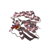
| Deposited unit | 
| ||||||||
|---|---|---|---|---|---|---|---|---|---|
| 1 |
| ||||||||
| Unit cell |
|
-Components
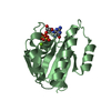
| #1: Protein | Mass: 19793.441 Da / Num. of mol.: 1 Source method: isolated from a genetically manipulated source Source: (gene. exp.) Homo sapiens (human) / Gene: RAB6B / Plasmid: pET28a / Species (production host): Escherichia coli / Production host:  Escherichia coli BL21(DE3) (bacteria) / Strain (production host): BL21(DE3) Escherichia coli BL21(DE3) (bacteria) / Strain (production host): BL21(DE3)References: UniProt: Q9NRW1, Hydrolases; Acting on acid anhydrides; In phosphorus-containing anhydrides |
|---|---|
| #2: Chemical | ChemComp-MG /   Mass: 24.305 Da / Num. of mol.: 1 / Source method: obtained synthetically / Formula: Mg Mass: 24.305 Da / Num. of mol.: 1 / Source method: obtained synthetically / Formula: Mg |
| #3: Chemical | ChemComp-NO3 / Nitrate  Mass: 62.005 Da / Num. of mol.: 1 / Source method: obtained synthetically / Formula: NO3 Mass: 62.005 Da / Num. of mol.: 1 / Source method: obtained synthetically / Formula: NO3 |
| #4: Chemical | ChemComp-GDP / Guanosine diphosphate  Type: RNA linking / Mass: 443.201 Da / Num. of mol.: 1 / Source method: obtained synthetically / Formula: C10H15N5O11P2 / Comment: GDP, energy-carrying molecule*YM Type: RNA linking / Mass: 443.201 Da / Num. of mol.: 1 / Source method: obtained synthetically / Formula: C10H15N5O11P2 / Comment: GDP, energy-carrying molecule*YM |
| #5: Water | ChemComp-HOH / Water Mass: 18.015 Da / Num. of mol.: 79 / Source method: isolated from a natural source / Formula: H2O Mass: 18.015 Da / Num. of mol.: 79 / Source method: isolated from a natural source / Formula: H2O |
-Experimental details
-Experiment
| Experiment | Method: X-RAY DIFFRACTION / Number of used crystals: 1 |
|---|
- Sample preparation
| Crystal | Density Matthews: 1.85 Å3/Da / Density % sol: 33.65 % |
|---|---|
| Crystal grow | Temperature: 292.15 K / Method: vapor diffusion, hanging drop / pH: 5 Details: 0.1M sodium acetate pH 5.0, 0.15M ammonium nitrate, 20%-23% PEG3350, VAPOR DIFFUSION, HANGING DROP, temperature 292.15K |
-Data collection
| Diffraction | Mean temperature: 100 K |
|---|---|
| Diffraction source | Source: SYNCHROTRON / Site: ESRF  / Beamline: ID14-2 / Wavelength: 0.933 Å / Beamline: ID14-2 / Wavelength: 0.933 Å |
| Detector | Type: ADSC QUANTUM 4 / Detector: CCD / Date: Jun 12, 2005 |
| Radiation | Protocol: SINGLE WAVELENGTH / Monochromatic (M) / Laue (L): M / Scattering type: x-ray |
| Radiation wavelength | Wavelength: 0.933 Å / Relative weight: 1 |
| Reflection | Resolution: 2.3→31.6 Å / Num. all: 6408 / Num. obs: 6366 / % possible obs: 91.6 % / Observed criterion σ(F): 2 / Observed criterion σ(I): 1 / Redundancy: 7.3 % / Rmerge(I) obs: 0.056 / Rsym value: 0.052 / Net I/σ(I): 10.2 |
| Reflection shell | Resolution: 2.3→2.42 Å / Redundancy: 7.5 % / Rmerge(I) obs: 0.076 / Mean I/σ(I) obs: 8.8 / Num. unique all: 984 / Rsym value: 0.07 / % possible all: 91.6 |
- Processing
| Software |
| ||||||||||||||||||||
|---|---|---|---|---|---|---|---|---|---|---|---|---|---|---|---|---|---|---|---|---|---|
| Refinement | Method to determine structure: Rab6 from Plasmodium falciparum (PDB code 1D5C) Resolution: 2.3→6 Å / Cross valid method: THROUGHOUT / σ(F): 2 / Stereochemistry target values: Engh & Huber
| ||||||||||||||||||||
| Refine analyze |
| ||||||||||||||||||||
| Refinement step | Cycle: LAST / Resolution: 2.3→6 Å
| ||||||||||||||||||||
| Refine LS restraints |
| ||||||||||||||||||||
| LS refinement shell | Resolution: 2.3→2.44 Å / Rfactor Rfree error: 0.028 /
|
