 Movie
Movie Controller

[English] 日本語
 Yorodumi
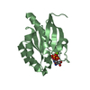
- PDB-2cxh: Crystal structure of probable ribosomal biogenesis protein from A... -
+ Open data

- Basic information
| Entry | Database: PDB / ID: 2cxh | ||||||
|---|---|---|---|---|---|---|---|
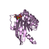
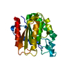
| Title | Crystal structure of probable ribosomal biogenesis protein from Aeropyrum pernix K1 | ||||||
 Components Components | Probable brix-domain ribosomal biogenesis protein | ||||||
 Keywords Keywords |  RNA BINDING PROTEIN / BRIX domain / 18S rRNA / IMP4 / U3 snoRNP / ribosomal biogenesis / RNA-binding / Structural Genomics / NPPSFA / National Project on Protein Structural and Functional Analyses / RIKEN Structural Genomics/Proteomics Initiative / RSGI RNA BINDING PROTEIN / BRIX domain / 18S rRNA / IMP4 / U3 snoRNP / ribosomal biogenesis / RNA-binding / Structural Genomics / NPPSFA / National Project on Protein Structural and Functional Analyses / RIKEN Structural Genomics/Proteomics Initiative / RSGI | ||||||
| Function / homology |  Function and homology information Function and homology information | ||||||
| Biological species |   Aeropyrum pernix (archaea) Aeropyrum pernix (archaea) | ||||||
| Method | X-RAY DIFFRACTION / SYNCHROTRON / SAD / Resolution: 1.8 Å | ||||||
 Authors Authors | Kawazoe, M. / Takemoto, C. / Hanawa-Suetsugu, K. / Kaminishi, T. / Terada, T. / Shirouzu, M. / Yokoyama, S. / RIKEN Structural Genomics/Proteomics Initiative (RSGI) | ||||||
 Citation Citation | Journal: To be Published Title: Crystal structure of probable ribosomal biogenesis protein from Aeropyrum pernix K1 Authors: Kawazoe, M. / Takemoto, C. / Hanawa-Suetsugu, K. / Kaminishi, T. / Terada, T. / Shirouzu, M. / Yokoyama, S. | ||||||
| History |
|
- Structure visualization
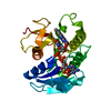
| Structure viewer | Molecule: MolmilJmol/JSmol |
|---|
- Downloads & links
-Download
| PDBx/mmCIF format | 2cxh.cif.gz | 47.9 KB | Display | PDBx/mmCIF format |
|---|---|---|---|---|
| PDB format | pdb2cxh.ent.gz | 36.6 KB | Display | PDB format |
| PDBx/mmJSON format | 2cxh.json.gz | Tree view | PDBx/mmJSON format | |
| Others |  Other downloads Other downloads |
-Validation report
| Arichive directory | https://data.pdbj.org/pub/pdb/validation_reports/cx/2cxhftp://data.pdbj.org/pub/pdb/validation_reports/cx/2cxh | HTTPS FTP |
|---|
-Related structure data
| Similar structure data | |
|---|---|
| Other databases |


-Links
 PDBj
PDBj- Assembly
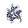
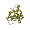
| Deposited unit | 
| ||||||||
|---|---|---|---|---|---|---|---|---|---|
| 1 |
| ||||||||
| Unit cell |
|
-Components
| #1: Protein | Mass: 23774.678 Da / Num. of mol.: 1 Source method: isolated from a genetically manipulated source Source: (gene. exp.) Aeropyrum pernix (archaea) / Strain: K1 / Gene: APE1443 / Plasmid: pET15b / Production host:  Escherichia coli (E. coli) / Strain (production host): B834 (DE3) pRARE / References: UniProt: Q9YC08 Escherichia coli (E. coli) / Strain (production host): B834 (DE3) pRARE / References: UniProt: Q9YC08 |
|---|---|
| #2: Water | ChemComp-HOH / Water Mass: 18.015 Da / Num. of mol.: 150 / Source method: isolated from a natural source / Formula: H2O Mass: 18.015 Da / Num. of mol.: 150 / Source method: isolated from a natural source / Formula: H2O |
-Experimental details
-Experiment
| Experiment | Method: X-RAY DIFFRACTION / Number of used crystals: 1 |
|---|
- Sample preparation
| Crystal | Density Matthews: 2.26692 Å3/Da / Density % sol: 45.741364 % |
|---|---|
| Crystal grow | Temperature: 293 K / Method: vapor diffusion, sitting drop Details: 0.01M Tris-HCl pH 8.0, 0.15M NaCl, 0.001M DTT, 0.05M tri-sodium citrate pH 5.6, 10% v/v 2-propanol, 10% w/v polyethylene glycol 4000, VAPOR DIFFUSION, SITTING DROP, temperature 293K |
-Data collection
| Diffraction | Mean temperature: 100 K |
|---|---|
| Diffraction source | Source: SYNCHROTRON / Site: Photon Factory  / Beamline: AR-NW12A / Wavelength: 0.9792 Å / Beamline: AR-NW12A / Wavelength: 0.9792 Å |
| Detector | Type: ADSC QUANTUM 210 / Detector: CCD / Date: Apr 15, 2005 / Details: mirrors |
| Radiation | Monochromator: Si 111 CHANNEL / Protocol: SINGLE WAVELENGTH / Monochromatic (M) / Laue (L): M / Scattering type: x-ray |
| Radiation wavelength | Wavelength: 0.9792 Å / Relative weight: 1 |
| Reflection | Resolution: 1.697→37.796 Å / Num. obs: 22973 / % possible obs: 93.6 % / Observed criterion σ(I): -3 / Redundancy: 8.36835 % / Biso Wilson estimate: 11 Å2 / Rsym value: 0.09 / Net I/σ(I): 23.616 |
| Reflection shell | Resolution: 1.7→1.76 Å / Redundancy: 8.1 % / Mean I/σ(I) obs: 2.5 / Num. unique all: 22973 / Rsym value: 0.608 / % possible all: 95.4 |
- Processing
| Software |
| ||||||||||||||||||||||||||||||||||||||||||||||||||||||||||||||||||||||||||||||||
|---|---|---|---|---|---|---|---|---|---|---|---|---|---|---|---|---|---|---|---|---|---|---|---|---|---|---|---|---|---|---|---|---|---|---|---|---|---|---|---|---|---|---|---|---|---|---|---|---|---|---|---|---|---|---|---|---|---|---|---|---|---|---|---|---|---|---|---|---|---|---|---|---|---|---|---|---|---|---|---|---|---|
| Refinement | Method to determine structure: SAD / Resolution: 1.8→37.71 Å / Rfactor Rfree error: 0.007 / Data cutoff high absF: 1210188.93 / Data cutoff low absF: 0 / Isotropic thermal model: RESTRAINED / Cross valid method: THROUGHOUT / σ(F): 0
| ||||||||||||||||||||||||||||||||||||||||||||||||||||||||||||||||||||||||||||||||
| Solvent computation | Solvent model: FLAT MODEL / Bsol: 38.244 Å2 / ksol: 0.36566 e/Å3 | ||||||||||||||||||||||||||||||||||||||||||||||||||||||||||||||||||||||||||||||||
| Displacement parameters | Biso mean: 19.9 Å2
| ||||||||||||||||||||||||||||||||||||||||||||||||||||||||||||||||||||||||||||||||
| Refine analyze |
| ||||||||||||||||||||||||||||||||||||||||||||||||||||||||||||||||||||||||||||||||
| Refinement step | Cycle: LAST / Resolution: 1.8→37.71 Å
| ||||||||||||||||||||||||||||||||||||||||||||||||||||||||||||||||||||||||||||||||
| Refine LS restraints |
| ||||||||||||||||||||||||||||||||||||||||||||||||||||||||||||||||||||||||||||||||
| LS refinement shell | Resolution: 1.8→1.91 Å / Rfactor Rfree error: 0.018 / Total num. of bins used: 6
| ||||||||||||||||||||||||||||||||||||||||||||||||||||||||||||||||||||||||||||||||
| Xplor file |
|
