 Movie
Movie Controller


+ Open data

- Basic information
| Entry | Database: PDB / ID: 7fpl | |||||||||||||||
|---|---|---|---|---|---|---|---|---|---|---|---|---|---|---|---|---|
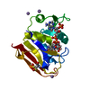
| Title | DHFR:NADP+:FOL complex at 280 K (crystal 1) | |||||||||||||||
 Components Components | Dihydrofolate reductase | |||||||||||||||
 Keywords Keywords | OXIDOREDUCTASE / Dihydrofolate Reductase | |||||||||||||||
| Function / homology |  Function and homology informationmethotrexate binding / dihydrofolic acid binding / response to methotrexate / dihydrofolate metabolic process / NADP+ binding / folic acid binding / folic acid metabolic process / glycine biosynthetic process / dihydrofolate reductase / dihydrofolate reductase activity ...methotrexate binding / dihydrofolic acid binding / response to methotrexate / dihydrofolate metabolic process / NADP+ binding / folic acid binding / folic acid metabolic process / glycine biosynthetic process / dihydrofolate reductase / dihydrofolate reductase activity / NADPH binding / tetrahydrofolate biosynthetic process / one-carbon metabolic process / NADP binding / response to xenobiotic stimulus / response to antibiotic / cytosol Function and homology informationmethotrexate binding / dihydrofolic acid binding / response to methotrexate / dihydrofolate metabolic process / NADP+ binding / folic acid binding / folic acid metabolic process / glycine biosynthetic process / dihydrofolate reductase / dihydrofolate reductase activity ...methotrexate binding / dihydrofolic acid binding / response to methotrexate / dihydrofolate metabolic process / NADP+ binding / folic acid binding / folic acid metabolic process / glycine biosynthetic process / dihydrofolate reductase / dihydrofolate reductase activity / NADPH binding / tetrahydrofolate biosynthetic process / one-carbon metabolic process / NADP binding / response to xenobiotic stimulus / response to antibiotic / cytosolSimilarity search - Function | |||||||||||||||
| Biological species |  Escherichia coli K-12 (bacteria) Escherichia coli K-12 (bacteria) | |||||||||||||||
| Method | X-RAY DIFFRACTION / SYNCHROTRON / FOURIER SYNTHESIS / Resolution: 1.17 Å | |||||||||||||||
 Authors Authors | Greisman, J.B. / Dalton, K.M. / Brookner, D.E. / Hekstra, D.R. | |||||||||||||||
| Funding support |  United States, 4items United States, 4items
| |||||||||||||||
 Citation Citation | Journal: To Be Published Title: DHFR:NADP+:FOL complex at 280 K Authors: Greisman, J.B. / Dalton, K.M. / Brookner, D.E. / Hekstra, D.R. | |||||||||||||||
| History |
|
- Structure visualization
| Structure viewer | Molecule: MolmilJmol/JSmol |
|---|
- Downloads & links
-Download
| PDBx/mmCIF format | 7fpl.cif.gz | 134.4 KB | Display | PDBx/mmCIF format |
|---|---|---|---|---|
| PDB format | pdb7fpl.ent.gz | 108.4 KB | Display | PDB format |
| PDBx/mmJSON format | 7fpl.json.gz | Tree view | PDBx/mmJSON format | |
| Others |  Other downloads Other downloads |
-Validation report
| Arichive directory | https://data.pdbj.org/pub/pdb/validation_reports/fp/7fplftp://data.pdbj.org/pub/pdb/validation_reports/fp/7fpl | HTTPS FTP |
|---|
-Group deposition
| ID | G_1002246 (6 entries) |
|---|---|
| Title | DHFR:NADP+:FOL complex at 280 K |
| Type | undefined |
| Description | DHFR:NADP+:FOL complex at 280 K |


-Related structure data
| Related structure data | 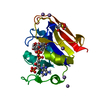 7lvcS S: Starting model for refinement |
|---|---|
| Similar structure data |
-Links
 PDBj
PDBj
- Assembly
| Deposited unit | 
| ||||||||||||
|---|---|---|---|---|---|---|---|---|---|---|---|---|---|
| 1 |
| ||||||||||||
| Unit cell |
|
-Components
| #1: Protein | Mass: 18051.338 Da / Num. of mol.: 1 Source method: isolated from a genetically manipulated source Source: (gene. exp.) Escherichia coli K-12 (bacteria) / Strain: K12 / Gene: folA, tmrA, b0048, JW0047 / Production host: Escherichia coli (E. coli) / References: UniProt: P0ABQ4, dihydrofolate reductase | ||||
|---|---|---|---|---|---|
| #2: Chemical | ChemComp-FOL / Folate  Mass: 441.397 Da / Num. of mol.: 1 / Source method: obtained synthetically / Formula: C19H19N7O6 / Feature type: SUBJECT OF INVESTIGATION Mass: 441.397 Da / Num. of mol.: 1 / Source method: obtained synthetically / Formula: C19H19N7O6 / Feature type: SUBJECT OF INVESTIGATION | ||||
| #3: Chemical | ChemComp-NAP / Nicotinamide adenine dinucleotide phosphate  Mass: 743.405 Da / Num. of mol.: 1 / Source method: obtained synthetically / Formula: C21H28N7O17P3 / Feature type: SUBJECT OF INVESTIGATION Mass: 743.405 Da / Num. of mol.: 1 / Source method: obtained synthetically / Formula: C21H28N7O17P3 / Feature type: SUBJECT OF INVESTIGATION | ||||
| #4: Chemical |   Mass: 54.938 Da / Num. of mol.: 3 / Source method: obtained synthetically / Formula: Mn Mass: 54.938 Da / Num. of mol.: 3 / Source method: obtained synthetically / Formula: Mn#5: Water | ChemComp-HOH / | Water Mass: 18.015 Da / Num. of mol.: 165 / Source method: isolated from a natural source / Formula: H2O Mass: 18.015 Da / Num. of mol.: 165 / Source method: isolated from a natural source / Formula: H2OHas ligand of interest | Y | |
-Experimental details
-Experiment
| Experiment | Method: X-RAY DIFFRACTION |
|---|
- Sample preparation
| Crystal | Density Matthews: 2.13 Å3/Da / Density % sol: 42.24 % |
|---|---|
| Crystal grow | Temperature: 277 K / Method: vapor diffusion, sitting drop / pH: 5.6 Details: 20 mM imadazole (pH 5.4-5.8), 16-21% PEG 400, 125 mM MnCl2 PH range: 5.4 - 5.8 |
-Data collection
| Diffraction | Mean temperature: 280 K / Serial crystal experiment: N |
|---|---|
| Diffraction source | Source: SYNCHROTRON / Site: SSRL / Beamline: BL12-1 / Wavelength: 0.826533 / Wavelength: 0.826533 Å |
| Detector | Type: DECTRIS EIGER2 S 16M / Detector: PIXEL / Date: Nov 20, 2021 |
| Radiation | Protocol: SINGLE WAVELENGTH / Scattering type: x-ray |
| Radiation wavelength | Wavelength: 0.826533 Å / Relative weight: 1 |
| Reflection | Resolution: 1.17→32.26 Å / Num. obs: 98434 / % possible obs: 97.8 % / Redundancy: 28.4 % / Biso Wilson estimate: 16.14 Å2 / CC1/2: 0.999 / Rmerge(I) obs: 0.285 / Rpim(I) all: 0.054 / Net I/σ(I): 18 |
| Reflection shell | Resolution: 1.17→1.19 Å / % possible obs: 96.7 % / Redundancy: 28.4 % / Rmerge(I) obs: 8.42 / Mean I/σ(I) obs: 0.7 / Num. unique obs: 4836 / CC1/2: 0.343 / Rpim(I) all: 1.586 |
- Processing
| Software |
| |||||||||||||||||||||||||||||||||||||||||||||||||||||||||||||||||||||||||||||||||||||||||||||||||||||||||||||||||||||||||||||||||||||||||||||||||||||||||||||||||||||||||||||||||||||||||||||||||||||||||||||||||||||||||
|---|---|---|---|---|---|---|---|---|---|---|---|---|---|---|---|---|---|---|---|---|---|---|---|---|---|---|---|---|---|---|---|---|---|---|---|---|---|---|---|---|---|---|---|---|---|---|---|---|---|---|---|---|---|---|---|---|---|---|---|---|---|---|---|---|---|---|---|---|---|---|---|---|---|---|---|---|---|---|---|---|---|---|---|---|---|---|---|---|---|---|---|---|---|---|---|---|---|---|---|---|---|---|---|---|---|---|---|---|---|---|---|---|---|---|---|---|---|---|---|---|---|---|---|---|---|---|---|---|---|---|---|---|---|---|---|---|---|---|---|---|---|---|---|---|---|---|---|---|---|---|---|---|---|---|---|---|---|---|---|---|---|---|---|---|---|---|---|---|---|---|---|---|---|---|---|---|---|---|---|---|---|---|---|---|---|---|---|---|---|---|---|---|---|---|---|---|---|---|---|---|---|---|---|---|---|---|---|---|---|---|---|---|---|---|---|---|---|---|
| Refinement | Method to determine structure: FOURIER SYNTHESIS Starting model: 7LVC Resolution: 1.17→22.75 Å / SU ML: 0.133 / Cross valid method: FREE R-VALUE / σ(F): 1.33 / Phase error: 17.8001 Stereochemistry target values: GeoStd + Monomer Library + CDL v1.2
| |||||||||||||||||||||||||||||||||||||||||||||||||||||||||||||||||||||||||||||||||||||||||||||||||||||||||||||||||||||||||||||||||||||||||||||||||||||||||||||||||||||||||||||||||||||||||||||||||||||||||||||||||||||||||
| Solvent computation | Shrinkage radii: 0.9 Å / VDW probe radii: 1.11 Å / Solvent model: FLAT BULK SOLVENT MODEL | |||||||||||||||||||||||||||||||||||||||||||||||||||||||||||||||||||||||||||||||||||||||||||||||||||||||||||||||||||||||||||||||||||||||||||||||||||||||||||||||||||||||||||||||||||||||||||||||||||||||||||||||||||||||||
| Displacement parameters | Biso mean: 21.99 Å2 | |||||||||||||||||||||||||||||||||||||||||||||||||||||||||||||||||||||||||||||||||||||||||||||||||||||||||||||||||||||||||||||||||||||||||||||||||||||||||||||||||||||||||||||||||||||||||||||||||||||||||||||||||||||||||
| Refinement step | Cycle: LAST / Resolution: 1.17→22.75 Å
| |||||||||||||||||||||||||||||||||||||||||||||||||||||||||||||||||||||||||||||||||||||||||||||||||||||||||||||||||||||||||||||||||||||||||||||||||||||||||||||||||||||||||||||||||||||||||||||||||||||||||||||||||||||||||
| Refine LS restraints |
| |||||||||||||||||||||||||||||||||||||||||||||||||||||||||||||||||||||||||||||||||||||||||||||||||||||||||||||||||||||||||||||||||||||||||||||||||||||||||||||||||||||||||||||||||||||||||||||||||||||||||||||||||||||||||
| LS refinement shell |
|
