 Movie
Movie Controller


+ Open data

- Basic information
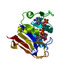
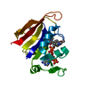
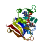
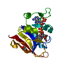
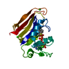
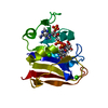
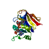
| Entry | Database: PDB / ID: 7lvc | ||||||||||||
|---|---|---|---|---|---|---|---|---|---|---|---|---|---|
| Title | E. coli DHFR by Native Mn,P,S-SAD at Room Temperature | ||||||||||||
 Components Components | Dihydrofolate reductase | ||||||||||||
 Keywords Keywords | OXIDOREDUCTASE / Dihydrofolate Reductase | ||||||||||||
| Function / homology |  Function and homology informationmethotrexate binding / dihydrofolic acid binding / response to methotrexate / NADP+ binding / folic acid binding / dihydrofolate metabolic process / glycine biosynthetic process / dihydrofolate reductase / folic acid metabolic process / dihydrofolate reductase activity ...methotrexate binding / dihydrofolic acid binding / response to methotrexate / NADP+ binding / folic acid binding / dihydrofolate metabolic process / glycine biosynthetic process / dihydrofolate reductase / folic acid metabolic process / dihydrofolate reductase activity / NADPH binding / tetrahydrofolate biosynthetic process / one-carbon metabolic process / NADP binding / response to xenobiotic stimulus / response to antibiotic / cytosol Function and homology informationmethotrexate binding / dihydrofolic acid binding / response to methotrexate / NADP+ binding / folic acid binding / dihydrofolate metabolic process / glycine biosynthetic process / dihydrofolate reductase / folic acid metabolic process / dihydrofolate reductase activity ...methotrexate binding / dihydrofolic acid binding / response to methotrexate / NADP+ binding / folic acid binding / dihydrofolate metabolic process / glycine biosynthetic process / dihydrofolate reductase / folic acid metabolic process / dihydrofolate reductase activity / NADPH binding / tetrahydrofolate biosynthetic process / one-carbon metabolic process / NADP binding / response to xenobiotic stimulus / response to antibiotic / cytosolSimilarity search - Function | ||||||||||||
| Biological species |  Escherichia coli (E. coli) Escherichia coli (E. coli) | ||||||||||||
| Method | X-RAY DIFFRACTION / SYNCHROTRON / SAD / Resolution: 1.7 Å | ||||||||||||
 Authors Authors | Greisman, J.B. / Dalton, K.M. / Hekstra, D.R. | ||||||||||||
| Funding support |  United States, 3items United States, 3items
| ||||||||||||
 Citation Citation | Journal: Acta Crystallogr D Struct Biol / Year: 2022 Title: Native SAD phasing at room temperature. Authors: Greisman, J.B. / Dalton, K.M. / Sheehan, C.J. / Klureza, M.A. / Kurinov, I. / Hekstra, D.R. #1: Journal: Acta Crystallogr.,Sect.DTitle: Native SAD phasing at room temperature Authors: Greisman, J.B. / Dalton, K.M. / Sheehan, C.J. / Klureza, M.A. / Kurinov, I. / Hekstra, D.R. | ||||||||||||
| History |
|
- Structure visualization
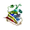
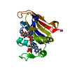
| Structure viewer | Molecule: MolmilJmol/JSmol |
|---|
- Downloads & links
-Download
| PDBx/mmCIF format | 7lvc.cif.gz | 129 KB | Display | PDBx/mmCIF format |
|---|---|---|---|---|
| PDB format | pdb7lvc.ent.gz | 95.4 KB | Display | PDB format |
| PDBx/mmJSON format | 7lvc.json.gz | Tree view | PDBx/mmJSON format | |
| Others |  Other downloads Other downloads |
-Validation report
| Arichive directory | https://data.pdbj.org/pub/pdb/validation_reports/lv/7lvcftp://data.pdbj.org/pub/pdb/validation_reports/lv/7lvc | HTTPS FTP |
|---|
-Related structure data
| Related structure data | 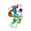 7l84C  7mm1C  7rinC C: citing same article ( |
|---|---|
| Similar structure data | |
| Experimental dataset #1 | Data reference: 10.15785/SBGRID/821 / Data set type: diffraction image data |

-Links
 PDBj
PDBj
- Assembly
| Deposited unit | 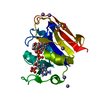
| ||||||||||||
|---|---|---|---|---|---|---|---|---|---|---|---|---|---|
| 1 |
| ||||||||||||
| Unit cell |
|
-Components
| #1: Protein | Mass: 18051.338 Da / Num. of mol.: 1 Source method: isolated from a genetically manipulated source Source: (gene. exp.) Escherichia coli (strain K12) (bacteria)Strain: K12 / Gene: folA, tmrA, b0048, JW0047 / Production host: Escherichia coli (E. coli) / References: UniProt: P0ABQ4, dihydrofolate reductase | ||||
|---|---|---|---|---|---|
| #2: Chemical | ChemComp-FOL / Folate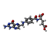  Mass: 441.397 Da / Num. of mol.: 1 / Source method: obtained synthetically / Formula: C19H19N7O6 Mass: 441.397 Da / Num. of mol.: 1 / Source method: obtained synthetically / Formula: C19H19N7O6 | ||||
| #3: Chemical | ChemComp-NAP / Nicotinamide adenine dinucleotide phosphate  Mass: 743.405 Da / Num. of mol.: 1 / Source method: obtained synthetically / Formula: C21H28N7O17P3 Mass: 743.405 Da / Num. of mol.: 1 / Source method: obtained synthetically / Formula: C21H28N7O17P3 | ||||
| #4: Chemical | ChemComp-MN /   Mass: 54.938 Da / Num. of mol.: 5 / Source method: obtained synthetically / Formula: Mn Mass: 54.938 Da / Num. of mol.: 5 / Source method: obtained synthetically / Formula: Mn#5: Water | ChemComp-HOH / | Water Mass: 18.015 Da / Num. of mol.: 106 / Source method: isolated from a natural source / Formula: H2O Mass: 18.015 Da / Num. of mol.: 106 / Source method: isolated from a natural source / Formula: H2OHas ligand of interest | N | |
-Experimental details
-Experiment
| Experiment | Method: X-RAY DIFFRACTION / Number of used crystals: 1 |
|---|
- Sample preparation
| Crystal | Density Matthews: 2.14 Å3/Da / Density % sol: 42.6 % |
|---|---|
| Crystal grow | Temperature: 277 K / Method: vapor diffusion, sitting drop Details: 20 mM imadazole (pH 5.4-5.8), 16-21% PEG 400, 125 mM MnCl2 PH range: 5.4 - 5.8 |
-Data collection
| Diffraction | Mean temperature: 295 K / Ambient temp details: ambient / Serial crystal experiment: N |
|---|---|
| Diffraction source | Source: SYNCHROTRON / Site: APS / Beamline: 24-ID-C / Wavelength: 1.892 Å |
| Detector | Type: DECTRIS PILATUS 6M-F / Detector: PIXEL / Date: Jul 17, 2019 |
| Radiation | Monochromator: M / Protocol: SINGLE WAVELENGTH / Monochromatic (M) / Laue (L): M / Scattering type: x-ray |
| Radiation wavelength | Wavelength: 1.892 Å / Relative weight: 1 |
| Reflection | Resolution: 1.7→49.52 Å / Num. obs: 27966 / % possible obs: 85.52 % / Redundancy: 29.4 % / Biso Wilson estimate: 12.78 Å2 / CC1/2: 0.998 / CC star: 1 / Rmerge(I) obs: 0.1012 / Rpim(I) all: 0.01671 / Rrim(I) all: 0.1026 / Net I/σ(I): 30.18 |
| Reflection shell | Resolution: 1.7→1.77 Å / Redundancy: 4.5 % / Rmerge(I) obs: 0.2227 / Mean I/σ(I) obs: 4.42 / Num. unique obs: 720 / CC1/2: 0.945 / CC star: 0.986 / Rpim(I) all: 0.1143 / Rrim(I) all: 0.2512 / % possible all: 22.11 |
- Processing
| Software |
| |||||||||||||||||||||||||||||||||||||||||||||||||||||||||||||||||||||||||||||
|---|---|---|---|---|---|---|---|---|---|---|---|---|---|---|---|---|---|---|---|---|---|---|---|---|---|---|---|---|---|---|---|---|---|---|---|---|---|---|---|---|---|---|---|---|---|---|---|---|---|---|---|---|---|---|---|---|---|---|---|---|---|---|---|---|---|---|---|---|---|---|---|---|---|---|---|---|---|---|
| Refinement | Method to determine structure: SAD / Resolution: 1.7→49.52 Å / SU ML: 0.1196 / Cross valid method: FREE R-VALUE / σ(F): 1.4 / Phase error: 16.4225 Stereochemistry target values: GeoStd + Monomer Library + CDL v1.2
| |||||||||||||||||||||||||||||||||||||||||||||||||||||||||||||||||||||||||||||
| Solvent computation | Shrinkage radii: 0.9 Å / VDW probe radii: 1.11 Å / Solvent model: FLAT BULK SOLVENT MODEL | |||||||||||||||||||||||||||||||||||||||||||||||||||||||||||||||||||||||||||||
| Displacement parameters | Biso mean: 16.83 Å2 | |||||||||||||||||||||||||||||||||||||||||||||||||||||||||||||||||||||||||||||
| Refinement step | Cycle: LAST / Resolution: 1.7→49.52 Å
| |||||||||||||||||||||||||||||||||||||||||||||||||||||||||||||||||||||||||||||
| Refine LS restraints |
| |||||||||||||||||||||||||||||||||||||||||||||||||||||||||||||||||||||||||||||
| LS refinement shell |
| |||||||||||||||||||||||||||||||||||||||||||||||||||||||||||||||||||||||||||||
| Refinement TLS params. | Method: refined / Origin x: 23.575962697 Å / Origin y: 21.4047757535 Å / Origin z: 35.5768107525 Å
| |||||||||||||||||||||||||||||||||||||||||||||||||||||||||||||||||||||||||||||
| Refinement TLS group | Selection details: all |
