 Movie
Movie Controller


+ Open data

- Basic information
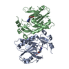
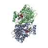
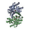
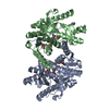
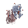
| Entry | Database: PDB / ID: 7f6j | ||||||
|---|---|---|---|---|---|---|---|
| Title | Crystal structure of the PDZD8 coiled-coil domain - Rab7 complex | ||||||
 Components Components |
| ||||||
 Keywords Keywords | LIPID TRANSPORT /  complex / PDZD8 / Rab7 / GTP / coiled-coil complex / PDZD8 / Rab7 / GTP / coiled-coil | ||||||
| Function / homology |  Function and homology information Function and homology informationlipophagy / positive regulation of viral process / phagosome acidification / protein to membrane docking / epidermal growth factor catabolic process / negative regulation of intralumenal vesicle formation / alveolar lamellar body / negative regulation of exosomal secretion / Suppression of autophagy / phagosome-lysosome fusion ...lipophagy / positive regulation of viral process / phagosome acidification / protein to membrane docking / epidermal growth factor catabolic process / negative regulation of intralumenal vesicle formation / alveolar lamellar body / negative regulation of exosomal secretion / Suppression of autophagy / phagosome-lysosome fusion / phagosome maturation / mitochondrial calcium ion homeostasis / establishment of vesicle localization / mitochondria-associated endoplasmic reticulum membrane contact site / retromer complex binding / endosome to plasma membrane protein transport / melanosome membrane / phagophore assembly site membrane / protein targeting to lysosome / RAB geranylgeranylation / early endosome to late endosome transport / positive regulation of exosomal secretion / RAB GEFs exchange GTP for GDP on RABs / RHOF GTPase cycle / RHOD GTPase cycle / regulation of cell morphogenesis / retrograde transport, endosome to Golgi / TBC/RABGAPs / lipid transport / mitochondrion-endoplasmic reticulum membrane tethering / endosome to lysosome transport / RHOJ GTPase cycle / RHOQ GTPase cycle / autophagosome membrane / RHOH GTPase cycle / CDC42 GTPase cycle / autophagosome assembly / RHOG GTPase cycle / viral release from host cell / intracellular transport / RAC2 GTPase cycle / RAC3 GTPase cycle / lipid catabolic process / bone resorption / phagocytic vesicle / Prevention of phagosomal-lysosomal fusion / cytoskeleton organization / RAC1 GTPase cycle / MHC class II antigen presentation / lipid droplet / small monomeric GTPase / G protein activity / secretory granule membrane / mitochondrial membrane / response to bacterium / synaptic vesicle membrane / small GTPase binding / endocytosis / positive regulation of protein catabolic process / phagocytic vesicle membrane / GDP binding / late endosome / protein transport / late endosome membrane / membrane => GO:0016020 / lysosome / endosome membrane / intracellular signal transduction / lysosomal membrane / GTPase activity / lipid binding / Neutrophil degranulation / endoplasmic reticulum membrane / GTP binding / Golgi apparatus / mitochondrion / extracellular exosome / membrane / metal ion binding / plasma membrane / cytosolSimilarity search - Function | ||||||
| Biological species |  Homo sapiens (human) Homo sapiens (human) | ||||||
| Method | X-RAY DIFFRACTION / SYNCHROTRON / MOLECULAR REPLACEMENT / Resolution: 2.1 Å | ||||||
 Authors Authors | Khan, H. / Chen, L. / Tan, L. / Im, Y.J. | ||||||
| Funding support |  Korea, Republic Of, 1items Korea, Republic Of, 1items
| ||||||
 Citation Citation | Journal: Sci Rep / Year: 2021 Title: Structural basis of human PDZD8-Rab7 interaction for the ER-late endosome tethering. Authors: Khan, H. / Chen, L. / Tan, L. / Im, Y.J. | ||||||
| History |
|
- Structure visualization
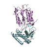
| Structure viewer | Molecule: MolmilJmol/JSmol |
|---|
- Downloads & links
-Download
| PDBx/mmCIF format | 7f6j.cif.gz | 120.9 KB | Display | PDBx/mmCIF format |
|---|---|---|---|---|
| PDB format | pdb7f6j.ent.gz | 82.4 KB | Display | PDB format |
| PDBx/mmJSON format | 7f6j.json.gz | Tree view | PDBx/mmJSON format | |
| Others |  Other downloads Other downloads |
-Validation report
| Arichive directory | https://data.pdbj.org/pub/pdb/validation_reports/f6/7f6jftp://data.pdbj.org/pub/pdb/validation_reports/f6/7f6j | HTTPS FTP |
|---|
-Related structure data
| Related structure data | 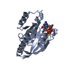 1t91S S: Starting model for refinement |
|---|---|
| Similar structure data |


-Links
 PDBj
PDBj


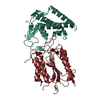
- Assembly
| Deposited unit | 
| ||||||||||||
|---|---|---|---|---|---|---|---|---|---|---|---|---|---|
| 1 |
| ||||||||||||
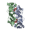
| Unit cell |
|
-Components
| #1: Protein | Mass: 22649.750 Da / Num. of mol.: 2 / Mutation: Q67L Source method: isolated from a genetically manipulated source Details: GTP-bound Rab7 Q67L / Source: (gene. exp.) Homo sapiens (human) / Gene: RAB7A, RAB7 / Plasmid: pHIS-2 / Details (production host): The N-terminal cleavable His-tag / Production host:  Escherichia coli BL21(DE3) (bacteria) / Strain (production host): BL21(DE3) / References: UniProt: P51149, small monomeric GTPase Escherichia coli BL21(DE3) (bacteria) / Strain (production host): BL21(DE3) / References: UniProt: P51149, small monomeric GTPase#2: Protein | | Mass: 15086.063 Da / Num. of mol.: 1 Source method: isolated from a genetically manipulated source Details: The coiled-coil domain of PDZD8 / Source: (gene. exp.) Homo sapiens (human) / Gene: PDZD8, PDZK8 / Plasmid: pHIS-2 / Details (production host): The N-terminal cleavable his-tag / Production host: Escherichia coli BL21(DE3) (bacteria) / Strain (production host): BL21(DE3) / References: UniProt: Q8NEN9#3: Chemical |   Mass: 24.305 Da / Num. of mol.: 2 / Source method: obtained synthetically / Formula: Mg / Feature type: SUBJECT OF INVESTIGATION Mass: 24.305 Da / Num. of mol.: 2 / Source method: obtained synthetically / Formula: Mg / Feature type: SUBJECT OF INVESTIGATION#4: Chemical | Guanosine triphosphate  Mass: 523.180 Da / Num. of mol.: 2 / Source method: obtained synthetically / Formula: C10H16N5O14P3 / Feature type: SUBJECT OF INVESTIGATION / Comment: GTP, energy-carrying molecule*YM Mass: 523.180 Da / Num. of mol.: 2 / Source method: obtained synthetically / Formula: C10H16N5O14P3 / Feature type: SUBJECT OF INVESTIGATION / Comment: GTP, energy-carrying molecule*YM#5: Water | ChemComp-HOH / | Water Mass: 18.015 Da / Num. of mol.: 154 / Source method: isolated from a natural source / Formula: H2O Mass: 18.015 Da / Num. of mol.: 154 / Source method: isolated from a natural source / Formula: H2OHas ligand of interest | Y | |
|---|
-Experimental details
-Experiment
| Experiment | Method: X-RAY DIFFRACTION / Number of used crystals: 1 |
|---|
- Sample preparation
| Crystal | Density Matthews: 2.62 Å3/Da / Density % sol: 53.09 % |
|---|---|
| Crystal grow | Temperature: 295 K / Method: vapor diffusion, hanging drop / pH: 8 / Details: 0.1 M Tris-HCl pH 8.0, 25% PEG 1000, 0.1 M NaCl |
-Data collection
| Diffraction | Mean temperature: 110 K / Serial crystal experiment: N |
|---|---|
| Diffraction source | Source: SYNCHROTRON / Site: PAL/PLS / Beamline: 7A (6B, 6C1) / Wavelength: 0.9795 Å |
| Detector | Type: ADSC QUANTUM 270 / Detector: CCD / Date: Oct 30, 2020 |
| Radiation | Protocol: SINGLE WAVELENGTH / Monochromatic (M) / Laue (L): M / Scattering type: x-ray |
| Radiation wavelength | Wavelength: 0.9795 Å / Relative weight: 1 |
| Reflection | Resolution: 2.1→50 Å / Num. obs: 36020 / % possible obs: 99.7 % / Redundancy: 11 % / Biso Wilson estimate: 34.3 Å2 / CC1/2: 0.997 / CC star: 0.999 / Rrim(I) all: 0.027 / Net I/σ(I): 53.9 |
| Reflection shell | Resolution: 2.1→2.14 Å / Redundancy: 11.3 % / Mean I/σ(I) obs: 7.8 / Num. unique obs: 1773 / CC1/2: 0.938 / CC star: 0.984 / Rrim(I) all: 0.506 / % possible all: 100 |
- Processing
| Software |
| |||||||||||||||||||||||||||||||||||||||||||||||||||||||||||||||||||||||||||||||||||||||||||||||||||||||||
|---|---|---|---|---|---|---|---|---|---|---|---|---|---|---|---|---|---|---|---|---|---|---|---|---|---|---|---|---|---|---|---|---|---|---|---|---|---|---|---|---|---|---|---|---|---|---|---|---|---|---|---|---|---|---|---|---|---|---|---|---|---|---|---|---|---|---|---|---|---|---|---|---|---|---|---|---|---|---|---|---|---|---|---|---|---|---|---|---|---|---|---|---|---|---|---|---|---|---|---|---|---|---|---|---|---|---|
| Refinement | Method to determine structure: MOLECULAR REPLACEMENT Starting model: 1T91 Resolution: 2.1→29.77 Å / SU ML: 0.248 / Cross valid method: FREE R-VALUE / σ(F): 1.34 / Phase error: 27.1218 Stereochemistry target values: GeoStd + Monomer Library + CDL v1.2
| |||||||||||||||||||||||||||||||||||||||||||||||||||||||||||||||||||||||||||||||||||||||||||||||||||||||||
| Solvent computation | Shrinkage radii: 0.9 Å / VDW probe radii: 1.11 Å / Solvent model: FLAT BULK SOLVENT MODEL | |||||||||||||||||||||||||||||||||||||||||||||||||||||||||||||||||||||||||||||||||||||||||||||||||||||||||
| Displacement parameters | Biso mean: 43.16 Å2 | |||||||||||||||||||||||||||||||||||||||||||||||||||||||||||||||||||||||||||||||||||||||||||||||||||||||||
| Refinement step | Cycle: LAST / Resolution: 2.1→29.77 Å
| |||||||||||||||||||||||||||||||||||||||||||||||||||||||||||||||||||||||||||||||||||||||||||||||||||||||||
| Refine LS restraints |
| |||||||||||||||||||||||||||||||||||||||||||||||||||||||||||||||||||||||||||||||||||||||||||||||||||||||||
| LS refinement shell |
|
