 Movie
Movie Controller


+ Open data

- Basic information
| Entry | Database: PDB / ID: 5wly | ||||||||||||
|---|---|---|---|---|---|---|---|---|---|---|---|---|---|
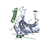
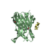
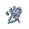
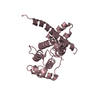
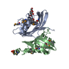
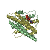
| Title | E. coli LpxH- 8 mutations | ||||||||||||
 Components Components | UDP-2,3-diacylglucosamine hydrolase UDP-2,3-diacylglucosamine diphosphatase UDP-2,3-diacylglucosamine diphosphatase | ||||||||||||
 Keywords Keywords | HYDROLASE / lipid A synthesis / open conformation / UDP-diacyl-glucosamine pyrophosphohydrolase / apha/beta-hydrolase | ||||||||||||
| Function / homology |  Function and homology informationUDP-2,3-diacylglucosamine diphosphatase / UDP-2,3-diacylglucosamine hydrolase activity / lipid A biosynthetic process / extrinsic component of plasma membrane / extrinsic component of cytoplasmic side of plasma membrane / manganese ion binding / cytoplasm Function and homology informationUDP-2,3-diacylglucosamine diphosphatase / UDP-2,3-diacylglucosamine hydrolase activity / lipid A biosynthetic process / extrinsic component of plasma membrane / extrinsic component of cytoplasmic side of plasma membrane / manganese ion binding / cytoplasmSimilarity search - Function | ||||||||||||
| Biological species |  Escherichia coli (E. coli) Escherichia coli (E. coli) | ||||||||||||
| Method | X-RAY DIFFRACTION / SYNCHROTRON / MOLECULAR REPLACEMENT / Resolution: 2 Å | ||||||||||||
 Authors Authors | Bohl, T.E. / Aihara, H. / Shi, K. / Lee, J.K. | ||||||||||||
| Funding support |  United States, 3items United States, 3items
| ||||||||||||
 Citation Citation | Journal: J. Biol. Chem. / Year: 2018 Title: The substrate-binding cap of the UDP-diacylglucosamine pyrophosphatase LpxH is highly flexible, enabling facile substrate binding and product release. Authors: Bohl, T.E. / Ieong, P. / Lee, J.K. / Lee, T. / Kankanala, J. / Shi, K. / Demir, O. / Kurahashi, K. / Amaro, R.E. / Wang, Z. / Aihara, H. | ||||||||||||
| History |
|
- Structure visualization
| Structure viewer | Molecule: MolmilJmol/JSmol |
|---|
- Downloads & links
-Download
| PDBx/mmCIF format | 5wly.cif.gz | 143.4 KB | Display | PDBx/mmCIF format |
|---|---|---|---|---|
| PDB format | pdb5wly.ent.gz | 111.1 KB | Display | PDB format |
| PDBx/mmJSON format | 5wly.json.gz | Tree view | PDBx/mmJSON format | |
| Others |  Other downloads Other downloads |
-Validation report
| Arichive directory | https://data.pdbj.org/pub/pdb/validation_reports/wl/5wlyftp://data.pdbj.org/pub/pdb/validation_reports/wl/5wly | HTTPS FTP |
|---|
-Related structure data
| Related structure data |  5b4aS S: Starting model for refinement |
|---|---|
| Similar structure data |


-Links
 PDBj
PDBj
- Assembly
| Deposited unit | 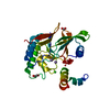
| ||||||||||||
|---|---|---|---|---|---|---|---|---|---|---|---|---|---|
| 1 |
| ||||||||||||
| 2 | 
| ||||||||||||
| Unit cell |
| ||||||||||||
| Components on special symmetry positions |
| ||||||||||||
| Details | monomer by gel filtration |
-Components
-Protein , 1 types, 1 molecules A
| #1: Protein | UDP-2,3-diacylglucosamine diphosphatase / UDP-2 / 3-diacylglucosamine diphosphatase Mass: 27731.977 Da / Num. of mol.: 1 Mutation: F141H, L142S, L146S, F147H, E14A, E15A, K161T, E162A Source method: isolated from a genetically manipulated source Source: (gene. exp.) Escherichia coli (E. coli) / Strain: DH5alphaGene: lpxH, AUQ13_20415, BK337_17315, BUE81_23120, EC3234A_5c00260, PGD_02789 Plasmid: pET24+ / Details (production host): short C-terminal His-tag / Production host: Escherichia coli BL21(DE3) (bacteria) / Strain (production host): BL-21 DE3References: UniProt: Q0P6L0, UniProt: P43341*PLUS, UDP-2,3-diacylglucosamine diphosphatase |
|---|
-Non-polymers , 5 types, 154 molecules 


| #2: Chemical |  Mass: 54.938 Da / Num. of mol.: 2 / Source method: obtained synthetically / Formula: Mn Mass: 54.938 Da / Num. of mol.: 2 / Source method: obtained synthetically / Formula: Mn#3: Chemical | ChemComp-CL / | Chloride Mass: 35.453 Da / Num. of mol.: 1 / Source method: obtained synthetically / Formula: Cl Mass: 35.453 Da / Num. of mol.: 1 / Source method: obtained synthetically / Formula: Cl#4: Chemical | ChemComp-FMT / | Formic acid Mass: 46.025 Da / Num. of mol.: 1 / Source method: obtained synthetically / Formula: CH2O2 Mass: 46.025 Da / Num. of mol.: 1 / Source method: obtained synthetically / Formula: CH2O2#5: Chemical | ChemComp-EDO / | Ethylene glycol Mass: 62.068 Da / Num. of mol.: 1 / Source method: obtained synthetically / Formula: C2H6O2 Mass: 62.068 Da / Num. of mol.: 1 / Source method: obtained synthetically / Formula: C2H6O2#6: Water | ChemComp-HOH / | WaterMass: 18.015 Da / Num. of mol.: 149 / Source method: isolated from a natural source / Formula: H2O |
|---|
-Experimental details
-Experiment
| Experiment | Method: X-RAY DIFFRACTION / Number of used crystals: 1 |
|---|
- Sample preparation
| Crystal | Density Matthews: 2.11 Å3/Da / Density % sol: 41.71 % / Description: parallelogram shaped plate crystals |
|---|---|
| Crystal grow | Temperature: 292 K / Method: vapor diffusion, hanging drop / pH: 8.2 Details: 7.1 g/L protein in 0.25 M NaCl, 10 mM Tris-HCl pH 7.4, 2.5 mM DTT, 1.25 mM MnCl2, and 20 mM reduced glutathione was combined 2:1 (protein to well solution) with 0.1 M Tris-HCl pH 8.2, 80 mM ...Details: 7.1 g/L protein in 0.25 M NaCl, 10 mM Tris-HCl pH 7.4, 2.5 mM DTT, 1.25 mM MnCl2, and 20 mM reduced glutathione was combined 2:1 (protein to well solution) with 0.1 M Tris-HCl pH 8.2, 80 mM magnesium formate, and 5% 2-propanol |
-Data collection
| Diffraction | Mean temperature: 100 K |
|---|---|
| Diffraction source | Source: SYNCHROTRON / Site: APS / Beamline: 24-ID-E / Wavelength: 0.97918 Å |
| Detector | Type: ADSC QUANTUM 315 / Detector: CCD / Date: Aug 22, 2016 |
| Radiation | Protocol: SINGLE WAVELENGTH / Monochromatic (M) / Laue (L): M / Scattering type: x-ray |
| Radiation wavelength | Wavelength: 0.97918 Å / Relative weight: 1 |
| Reflection | Resolution: 2→66.05 Å / Num. obs: 16046 / % possible obs: 98.6 % / Redundancy: 6 % / CC1/2: 0.994 / Rmerge(I) obs: 0.179 / Rpim(I) all: 0.078 / Net I/σ(I): 7.7 |
| Reflection shell | Resolution: 2→2.06 Å / Redundancy: 5.9 % / Rmerge(I) obs: 1.973 / Mean I/σ(I) obs: 0.8 / Num. unique obs: 1090 / CC1/2: 0.279 / Rpim(I) all: 0.867 / % possible all: 91 |
- Processing
| Software |
| ||||||||||||||||||
|---|---|---|---|---|---|---|---|---|---|---|---|---|---|---|---|---|---|---|---|
| Refinement | Method to determine structure: MOLECULAR REPLACEMENT Starting model: 5B4A Resolution: 2→45.23 Å / SU ML: 0.25 / Cross valid method: FREE R-VALUE / Phase error: 26.93
| ||||||||||||||||||
| Solvent computation | Shrinkage radii: 0.9 Å / VDW probe radii: 1.11 Å | ||||||||||||||||||
| Displacement parameters | Biso mean: 42.45 Å2 | ||||||||||||||||||
| Refinement step | Cycle: LAST / Resolution: 2→45.23 Å
| ||||||||||||||||||
| LS refinement shell | Resolution: 2→2.16 Å
|
