 Movie
Movie Controller


+ Open data

- Basic information
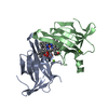
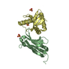
| Entry | Database: PDB / ID: 7f2j | |||||||||
|---|---|---|---|---|---|---|---|---|---|---|
| Title | Crystal structure of AtFKBP53 FKBD in complex with rapamycin | |||||||||
 Components Components | Peptidyl-prolyl cis-trans isomerase FKBP53 | |||||||||
 Keywords Keywords |  ISOMERASE / FKBP / rapamycin / dimerization ISOMERASE / FKBP / rapamycin / dimerization | |||||||||
| Function / homology |  Function and homology informationpeptidylprolyl isomerase / peptidyl-prolyl cis-trans isomerase activity / nucleosome assembly / histone binding / nucleolus / nucleus Function and homology informationpeptidylprolyl isomerase / peptidyl-prolyl cis-trans isomerase activity / nucleosome assembly / histone binding / nucleolus / nucleusSimilarity search - Function | |||||||||
| Biological species |  Arabidopsis thaliana (thale cress) Arabidopsis thaliana (thale cress) | |||||||||
| Method | X-RAY DIFFRACTION / SYNCHROTRON / MOLECULAR REPLACEMENT / Resolution: 1.6 Å | |||||||||
 Authors Authors | Singh, A.K. / Saharan, K. / Vasudevan, D. | |||||||||
| Funding support |  India, 2items India, 2items
| |||||||||
 Citation Citation | Journal: Int.J.Biol.Macromol. / Year: 2022 Title: Crystal packing reveals rapamycin-mediated homodimerization of an FK506-binding domain. Authors: Singh, A.K. / Saharan, K. / Baral, S. / Luan, S. / Vasudevan, D. | |||||||||
| History |
|
- Structure visualization
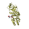
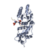
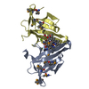
| Structure viewer | Molecule: MolmilJmol/JSmol |
|---|
- Downloads & links
-Download
| PDBx/mmCIF format | 7f2j.cif.gz | 67.1 KB | Display | PDBx/mmCIF format |
|---|---|---|---|---|
| PDB format | pdb7f2j.ent.gz | 47.2 KB | Display | PDB format |
| PDBx/mmJSON format | 7f2j.json.gz | Tree view | PDBx/mmJSON format | |
| Others |  Other downloads Other downloads |
-Validation report
| Arichive directory | https://data.pdbj.org/pub/pdb/validation_reports/f2/7f2jftp://data.pdbj.org/pub/pdb/validation_reports/f2/7f2j | HTTPS FTP |
|---|
-Related structure data
| Related structure data | 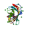 6j2mS S: Starting model for refinement |
|---|---|
| Similar structure data |
-Links
 PDBj
PDBj

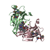
- Assembly
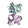
| Deposited unit | 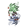
| ||||||||
|---|---|---|---|---|---|---|---|---|---|
| 1 |
| ||||||||
| Unit cell |
|
-Components
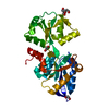
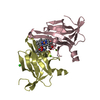
| #1: Protein | Mass: 13648.736 Da / Num. of mol.: 2 / Fragment: C-terminal domain Source method: isolated from a genetically manipulated source Source: (gene. exp.) Arabidopsis thaliana (thale cress) / Gene: FKBP53, At4g25340, T30C3_20 / Plasmid: pET22b / Production host:  Escherichia coli BL21(DE3) (bacteria) / References: UniProt: Q93ZG9, peptidylprolyl isomerase Escherichia coli BL21(DE3) (bacteria) / References: UniProt: Q93ZG9, peptidylprolyl isomerase#2: Chemical | Sirolimus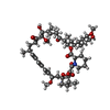  Mass: 914.172 Da / Num. of mol.: 2 / Source method: obtained synthetically / Formula: C51H79NO13 / Feature type: SUBJECT OF INVESTIGATION / Comment: immunosuppressant, antibiotic*YM Mass: 914.172 Da / Num. of mol.: 2 / Source method: obtained synthetically / Formula: C51H79NO13 / Feature type: SUBJECT OF INVESTIGATION / Comment: immunosuppressant, antibiotic*YM#3: Chemical | ChemComp-SO4 / Sulfate  Mass: 96.063 Da / Num. of mol.: 5 / Source method: obtained synthetically / Formula: SO4 Mass: 96.063 Da / Num. of mol.: 5 / Source method: obtained synthetically / Formula: SO4#4: Chemical | ChemComp-PEG / | Diethylene glycol  Mass: 106.120 Da / Num. of mol.: 1 / Source method: obtained synthetically / Formula: C4H10O3 Mass: 106.120 Da / Num. of mol.: 1 / Source method: obtained synthetically / Formula: C4H10O3#5: Water | ChemComp-HOH / | Water Mass: 18.015 Da / Num. of mol.: 85 / Source method: isolated from a natural source / Formula: H2O Mass: 18.015 Da / Num. of mol.: 85 / Source method: isolated from a natural source / Formula: H2OHas ligand of interest | Y | |
|---|
-Experimental details
-Experiment
| Experiment | Method: X-RAY DIFFRACTION / Number of used crystals: 1 |
|---|
- Sample preparation
| Crystal | Density Matthews: 2.53 Å3/Da / Density % sol: 51.36 % / Description: rod-shaped |
|---|---|
| Crystal grow | Temperature: 291 K / Method: vapor diffusion, sitting drop / pH: 6.5 Details: 0.2 M Lithium sulfate monohydrate, 0.1 M Bis-Tris (pH 6.5), 25% w/v PEG 3350 |
-Data collection
| Diffraction | Mean temperature: 100 K / Serial crystal experiment: N |
|---|---|
| Diffraction source | Source: SYNCHROTRON / Site: ESRF  / Beamline: ID23-2 / Wavelength: 0.9677 Å / Beamline: ID23-2 / Wavelength: 0.9677 Å |
| Detector | Type: DECTRIS EIGER X 4M / Detector: PIXEL / Date: Dec 1, 2017 |
| Radiation | Protocol: SINGLE WAVELENGTH / Monochromatic (M) / Laue (L): M / Scattering type: x-ray |
| Radiation wavelength | Wavelength: 0.9677 Å / Relative weight: 1 |
| Reflection | Resolution: 1.6→47.01 Å / Num. obs: 32798 / % possible obs: 99.2 % / Redundancy: 4.4 % / Biso Wilson estimate: 19.1 Å2 / CC1/2: 0.998 / Rmerge(I) obs: 0.063 / Net I/σ(I): 11 |
| Reflection shell | Resolution: 1.6→1.65 Å / Redundancy: 4.3 % / Rmerge(I) obs: 0.541 / Mean I/σ(I) obs: 2.3 / Num. unique obs: 1594 / CC1/2: 0.743 / % possible all: 98.6 |
- Processing
| Software |
| ||||||||||||||||||||||||||||||||||||||||||||||||||||||||||||||||||||||||||||||||||||||||||||||||||||||||||||||||||||||||||||||||||||||||||||||||||||||||||||||||||||||||||||||||||||||
|---|---|---|---|---|---|---|---|---|---|---|---|---|---|---|---|---|---|---|---|---|---|---|---|---|---|---|---|---|---|---|---|---|---|---|---|---|---|---|---|---|---|---|---|---|---|---|---|---|---|---|---|---|---|---|---|---|---|---|---|---|---|---|---|---|---|---|---|---|---|---|---|---|---|---|---|---|---|---|---|---|---|---|---|---|---|---|---|---|---|---|---|---|---|---|---|---|---|---|---|---|---|---|---|---|---|---|---|---|---|---|---|---|---|---|---|---|---|---|---|---|---|---|---|---|---|---|---|---|---|---|---|---|---|---|---|---|---|---|---|---|---|---|---|---|---|---|---|---|---|---|---|---|---|---|---|---|---|---|---|---|---|---|---|---|---|---|---|---|---|---|---|---|---|---|---|---|---|---|---|---|---|---|---|
| Refinement | Method to determine structure: MOLECULAR REPLACEMENT Starting model: 6J2M Resolution: 1.6→47.01 Å / Cor.coef. Fo:Fc: 0.961 / Cor.coef. Fo:Fc free: 0.953 / SU B: 3.01 / SU ML: 0.058 / Cross valid method: THROUGHOUT / ESU R: 0.086 / ESU R Free: 0.087 / Stereochemistry target values: MAXIMUM LIKELIHOOD / Details: HYDROGENS HAVE BEEN ADDED IN THE RIDING POSITIONS
| ||||||||||||||||||||||||||||||||||||||||||||||||||||||||||||||||||||||||||||||||||||||||||||||||||||||||||||||||||||||||||||||||||||||||||||||||||||||||||||||||||||||||||||||||||||||
| Solvent computation | Ion probe radii: 1 Å / Shrinkage radii: 1 Å / VDW probe radii: 1.4 Å / Solvent model: MASK | ||||||||||||||||||||||||||||||||||||||||||||||||||||||||||||||||||||||||||||||||||||||||||||||||||||||||||||||||||||||||||||||||||||||||||||||||||||||||||||||||||||||||||||||||||||||
| Displacement parameters | Biso mean: 18.01 Å2
| ||||||||||||||||||||||||||||||||||||||||||||||||||||||||||||||||||||||||||||||||||||||||||||||||||||||||||||||||||||||||||||||||||||||||||||||||||||||||||||||||||||||||||||||||||||||
| Refinement step | Cycle: LAST / Resolution: 1.6→47.01 Å
| ||||||||||||||||||||||||||||||||||||||||||||||||||||||||||||||||||||||||||||||||||||||||||||||||||||||||||||||||||||||||||||||||||||||||||||||||||||||||||||||||||||||||||||||||||||||
| Refine LS restraints |
|