 Movie
Movie Controller


+ Open data

- Basic information
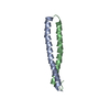
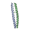
| Entry | Database: PDB / ID: 7ekn | ||||||
|---|---|---|---|---|---|---|---|
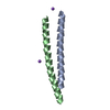
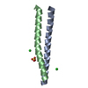
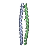
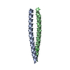
| Title | Crystal structure of AF10-ipep complex | ||||||
 Components Components |
| ||||||
 Keywords Keywords |  PROTEIN BINDING / inhibitor PROTEIN BINDING / inhibitor | ||||||
| Function / homology | nucleoplasm / cytosol / Protein AF-10 Function and homology information Function and homology information | ||||||
| Biological species |  Homo sapiens (human) Homo sapiens (human) | ||||||
| Method | X-RAY DIFFRACTION / SYNCHROTRON / MOLECULAR REPLACEMENT / Resolution: 2.14 Å | ||||||
 Authors Authors | Chen, S. / Zhou, Z. | ||||||
| Funding support |  China, 1items China, 1items
| ||||||
 Citation Citation | Journal: J Leukoc Biol / Year: 2021 Title: Structural characteristics of coiled-coil regions in AF10-DOT1L and AF10-inhibitory peptide complex. Authors: Zhou, Z. / Kang, S. / Huang, Z. / Zhou, Z. / Chen, S. | ||||||
| History |
|
- Structure visualization
| Structure viewer | Molecule: MolmilJmol/JSmol |
|---|
- Downloads & links
-Download
| PDBx/mmCIF format | 7ekn.cif.gz | 100.3 KB | Display | PDBx/mmCIF format |
|---|---|---|---|---|
| PDB format | pdb7ekn.ent.gz | 63.2 KB | Display | PDB format |
| PDBx/mmJSON format | 7ekn.json.gz | Tree view | PDBx/mmJSON format | |
| Others |  Other downloads Other downloads |
-Validation report
| Arichive directory | https://data.pdbj.org/pub/pdb/validation_reports/ek/7eknftp://data.pdbj.org/pub/pdb/validation_reports/ek/7ekn | HTTPS FTP |
|---|
-Related structure data
| Related structure data |  7edpSC S: Starting model for refinement C: citing same article ( |
|---|---|
| Similar structure data |


-Links
 PDBj
PDBj- Assembly
| Deposited unit | 
| ||||||||||
|---|---|---|---|---|---|---|---|---|---|---|---|
| 1 | 
| ||||||||||
| 2 | 
| ||||||||||
| 3 | 
| ||||||||||
| 4 | 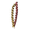
| ||||||||||
| Unit cell |
|
-Components
| #1: Protein/peptide | Mass: 5018.727 Da / Num. of mol.: 4 / Source method: obtained synthetically / Source: (synth.) Homo sapiens (human)#2: Protein/peptide | Mass: 5007.853 Da / Num. of mol.: 4 Source method: isolated from a genetically manipulated source Source: (gene. exp.) Homo sapiens (human) / Gene: MLLT10 / Production host:  Escherichia coli (E. coli) / References: UniProt: Q5JT35 Escherichia coli (E. coli) / References: UniProt: Q5JT35#3: Water | ChemComp-HOH / | Water Mass: 18.015 Da / Num. of mol.: 300 / Source method: isolated from a natural source / Formula: H2O Mass: 18.015 Da / Num. of mol.: 300 / Source method: isolated from a natural source / Formula: H2O |
|---|
-Experimental details
-Experiment
| Experiment | Method: X-RAY DIFFRACTION / Number of used crystals: 1 |
|---|
- Sample preparation
| Crystal | Density Matthews: 2.14 Å3/Da / Density % sol: 42.52 % |
|---|---|
| Crystal grow | Temperature: 293 K / Method: vapor diffusion, hanging drop / Details: PEG4000, Tris8.5, Sodium acetate |
-Data collection
| Diffraction | Mean temperature: 100 K / Serial crystal experiment: N |
|---|---|
| Diffraction source | Source: SYNCHROTRON / Site: SSRF / Beamline: BL18U1 / Wavelength: 0.9788 Å |
| Detector | Type: DECTRIS PILATUS3 6M / Detector: PIXEL / Date: Mar 1, 2015 |
| Radiation | Protocol: SINGLE WAVELENGTH / Monochromatic (M) / Laue (L): M / Scattering type: x-ray |
| Radiation wavelength | Wavelength: 0.9788 Å / Relative weight: 1 |
| Reflection | Resolution: 2.14→50 Å / Num. obs: 17862 / % possible obs: 97.5 % / Redundancy: 3.8 % / Biso Wilson estimate: 19.38 Å2 / Rmerge(I) obs: 0.129 / Rpim(I) all: 0.129 / Rrim(I) all: 0.182 / Net I/σ(I): 10.2 |
| Reflection shell | Resolution: 2.14→2.31 Å / Num. unique obs: 30000 / CC1/2: 0.648 |
- Processing
| Software |
| ||||||||||||||||||||||||||||||||||||||||||||||||||||||||||||||||||||||||||||||||||||||||||||||||||
|---|---|---|---|---|---|---|---|---|---|---|---|---|---|---|---|---|---|---|---|---|---|---|---|---|---|---|---|---|---|---|---|---|---|---|---|---|---|---|---|---|---|---|---|---|---|---|---|---|---|---|---|---|---|---|---|---|---|---|---|---|---|---|---|---|---|---|---|---|---|---|---|---|---|---|---|---|---|---|---|---|---|---|---|---|---|---|---|---|---|---|---|---|---|---|---|---|---|---|---|
| Refinement | Method to determine structure: MOLECULAR REPLACEMENT Starting model: 7EDP Resolution: 2.14→45.01 Å / SU ML: 0.2943 / Cross valid method: FREE R-VALUE / σ(F): 1.98 / Phase error: 26.5325 Stereochemistry target values: GeoStd + Monomer Library + CDL v1.2
| ||||||||||||||||||||||||||||||||||||||||||||||||||||||||||||||||||||||||||||||||||||||||||||||||||
| Solvent computation | Shrinkage radii: 0.9 Å / VDW probe radii: 1.11 Å / Solvent model: FLAT BULK SOLVENT MODEL | ||||||||||||||||||||||||||||||||||||||||||||||||||||||||||||||||||||||||||||||||||||||||||||||||||
| Displacement parameters | Biso mean: 23.98 Å2 | ||||||||||||||||||||||||||||||||||||||||||||||||||||||||||||||||||||||||||||||||||||||||||||||||||
| Refinement step | Cycle: LAST / Resolution: 2.14→45.01 Å
| ||||||||||||||||||||||||||||||||||||||||||||||||||||||||||||||||||||||||||||||||||||||||||||||||||
| Refine LS restraints |
| ||||||||||||||||||||||||||||||||||||||||||||||||||||||||||||||||||||||||||||||||||||||||||||||||||
| LS refinement shell |
|
