 Movie
Movie Controller


+ Open data

- Basic information
| Entry | Database: PDB / ID: 7e1y | ||||||
|---|---|---|---|---|---|---|---|
| Title | Staphylothermus marinus amylopullulanase -SmApu | ||||||
 Components Components | Glycoside hydrolase, family 57 | ||||||
 Keywords Keywords | HYDROLASE / amylopullulanase | ||||||
| Function / homology | Glycoside hydrolase family 57, N-terminal domain / Glycosyl hydrolase family 57 / Glycoside hydrolase 38, N-terminal domain superfamily / Glycoside hydrolase/deacetylase, beta/alpha-barrel / hydrolase activity / carbohydrate metabolic process / Glycoside hydrolase, family 57 Function and homology information Function and homology information | ||||||
| Biological species |  Staphylothermus marinus (archaea) Staphylothermus marinus (archaea) | ||||||
| Method | ELECTRON MICROSCOPY / single particle reconstruction / negative staining / cryo EM / Resolution: 2.9 Å | ||||||
 Authors Authors | Li, D. / Li, X. / Woo, E.-J. | ||||||
| Funding support |  China, 1items China, 1items
| ||||||
 Citation Citation | Journal: To Be Published Title: Staphylothermus marinus amylopullulanase -SmApu Authors: Li, D. / Li, X. | ||||||
| History |
|
- Structure visualization
| Movie |
Movie viewer |
|---|---|
| Structure viewer | Molecule: MolmilJmol/JSmol |
- Downloads & links
-Download
| PDBx/mmCIF format | 7e1y.cif.gz | 754.8 KB | Display | PDBx/mmCIF format |
|---|---|---|---|---|
| PDB format | pdb7e1y.ent.gz | 647.4 KB | Display | PDB format |
| PDBx/mmJSON format | 7e1y.json.gz | Tree view | PDBx/mmJSON format | |
| Others |  Other downloads Other downloads |
-Validation report
| Arichive directory | https://data.pdbj.org/pub/pdb/validation_reports/e1/7e1yftp://data.pdbj.org/pub/pdb/validation_reports/e1/7e1y | HTTPS FTP |
|---|
-Related structure data
| Related structure data | 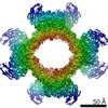 30946MC M: map data used to model this data C: citing same article ( |
|---|---|
| Similar structure data |

-Links
 PDBj
PDBj
- Assembly
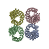
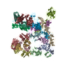
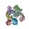
| Deposited unit | 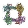
|
|---|---|
| 1 |
|
-Components
| #1: Protein | Mass: 75983.641 Da / Num. of mol.: 8 Source method: isolated from a genetically manipulated source Source: (gene. exp.) Staphylothermus marinus (strain ATCC 43588 / DSM 3639 / JCM 9404 / F1) (archaea)Strain: ATCC 43588 / DSM 3639 / JCM 9404 / F1 / Gene: Smar_1407 / Production host:  Escherichia coli BL21(DE3) (bacteria) / References: UniProt: A3DPD7 Escherichia coli BL21(DE3) (bacteria) / References: UniProt: A3DPD7 |
|---|
-Experimental details
-Experiment
| Experiment | Method: ELECTRON MICROSCOPY |
|---|---|
| EM experiment | Aggregation state: PARTICLE / 3D reconstruction method: single particle reconstruction |
- Sample preparation
| Component | Name: The octamer of glycoside hydrolase family 57. / Type: COMPLEX / Entity ID: all / Source: RECOMBINANT |
|---|---|
| Source (natural) | Organism: Staphylothermus marinus (archaea) |
| Source (recombinant) | Organism: Escherichia coli (E. coli) |
| Buffer solution | pH: 7.5 |
| Specimen | Conc.: 2 mg/ml / Embedding applied: NO / Shadowing applied: NO / Staining applied: YES / Vitrification applied: YES |
| EM staining | Type: NEGATIVE / Material: Uranyl acetate |
| Specimen support | Grid material: COPPER |
| Vitrification | Instrument: FEI VITROBOT MARK I / Cryogen name: ETHANE / Humidity: 100 % / Chamber temperature: 277 K |
- Electron microscopy imaging
| Experimental equipment |  Model: Titan Krios / Image courtesy: FEI Company |
|---|---|
| Microscopy | Model: FEI TITAN KRIOS |
| Electron gun | Electron source: FIELD EMISSION GUN / Accelerating voltage: 300 kV / Illumination mode: FLOOD BEAM |
| Electron lens | Mode: BRIGHT FIELDBright-field microscopy / Cs: 2.7 mm |
| Image recording | Average exposure time: 8 sec. / Electron dose: 64 e/Å2 / Detector mode: COUNTING / Film or detector model: GATAN K2 SUMMIT (4k x 4k) |
| Image scans | Movie frames/image: 40 |
- Processing
| Software | Name: PHENIX / Version: 1.18.2_3874: / Classification: refinement | ||||||||||||||||||||||||
|---|---|---|---|---|---|---|---|---|---|---|---|---|---|---|---|---|---|---|---|---|---|---|---|---|---|
| CTF correction | Type: PHASE FLIPPING AND AMPLITUDE CORRECTION | ||||||||||||||||||||||||
| 3D reconstruction | Resolution: 2.9 Å / Resolution method: FSC 0.143 CUT-OFF / Num. of particles: 272770 / Symmetry type: POINT | ||||||||||||||||||||||||
| Atomic model building | Protocol: AB INITIO MODEL / Space: REAL | ||||||||||||||||||||||||
| Refine LS restraints |
|
