 Movie
Movie Controller


+ Open data

- Basic information
| Entry | Database: EMDB / ID: EMD-30946 | |||||||||
|---|---|---|---|---|---|---|---|---|---|---|
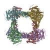
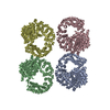
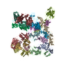
| Title | Staphylothermus marinus amylopullulanase -SmApu | |||||||||
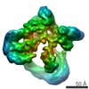
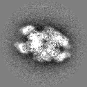
 Map data Map data | ||||||||||
 Sample Sample |
| |||||||||
| Function / homology | Glycoside hydrolase family 57, N-terminal domain / Glycosyl hydrolase family 57 /  Glycoside hydrolase 38, N-terminal domain superfamily / Glycoside hydrolase/deacetylase, beta/alpha-barrel / hydrolase activity / carbohydrate metabolic process / Glycoside hydrolase, family 57 Glycoside hydrolase 38, N-terminal domain superfamily / Glycoside hydrolase/deacetylase, beta/alpha-barrel / hydrolase activity / carbohydrate metabolic process / Glycoside hydrolase, family 57 Function and homology information Function and homology information | |||||||||
| Biological species |  Staphylothermus marinus (archaea) / Staphylothermus marinus (strain ATCC 43588 / DSM 3639 / JCM 9404 / F1) (archaea) Staphylothermus marinus (archaea) / Staphylothermus marinus (strain ATCC 43588 / DSM 3639 / JCM 9404 / F1) (archaea) | |||||||||
| Method | single particle reconstruction / cryo EM / negative staining / Resolution: 2.9 Å | |||||||||
 Authors Authors | Li D / Li X / Woo E-J | |||||||||
| Funding support |  China, 1 items China, 1 items
| |||||||||
 Citation Citation | Journal: To Be Published Title: Staphylothermus marinus amylopullulanase -SmApu Authors: Li D / Li X | |||||||||
| History |
|
- Structure visualization
| Movie |
Movie viewer |
|---|---|
| Structure viewer | EM map: SurfViewMolmilJmol/JSmol |
| Supplemental images |

- Downloads & links
-EMDB archive
| Map data | emd_30946.map.gz | 10.9 MB | EMDB map data format | |
|---|---|---|---|---|
| Header (meta data) | emd-30946-v30.xmlemd-30946.xml | 14.4 KB 14.4 KB | Display Display | EMDB header |
| FSC (resolution estimation) | emd_30946_fsc.xml | 10.7 KB | Display | FSC data file |
| Images |  emd_30946.png emd_30946.png | 90 KB | ||
| Masks | emd_30946_msk_1.map | 103 MB | Mask map | |
| Others | emd_30946_half_map_1.map.gzemd_30946_half_map_2.map.gz | 80.9 MB 80.8 MB | ||
| Archive directory |  http://ftp.pdbj.org/pub/emdb/structures/EMD-30946ftp://ftp.pdbj.org/pub/emdb/structures/EMD-30946 http://ftp.pdbj.org/pub/emdb/structures/EMD-30946ftp://ftp.pdbj.org/pub/emdb/structures/EMD-30946 | HTTPS FTP |
-Related structure data
| Related structure data |  7e1yMC M: atomic model generated by this map C: citing same article ( |
|---|---|
| Similar structure data |

-Links
| EMDB pages | EMDB (EBI/PDBe) / EMDataResource |
|---|---|
| Related items in Molecule of the Month |

-Map
| File | Download / File: emd_30946.map.gz / Format: CCP4 / Size: 103 MB / Type: IMAGE STORED AS FLOATING POINT NUMBER (4 BYTES) | ||||||||||||||||||||||||||||||||||||||||||||||||||||||||||||||||||||
|---|---|---|---|---|---|---|---|---|---|---|---|---|---|---|---|---|---|---|---|---|---|---|---|---|---|---|---|---|---|---|---|---|---|---|---|---|---|---|---|---|---|---|---|---|---|---|---|---|---|---|---|---|---|---|---|---|---|---|---|---|---|---|---|---|---|---|---|---|---|
| Voxel size | X=Y=Z: 1.014 Å | ||||||||||||||||||||||||||||||||||||||||||||||||||||||||||||||||||||
| Density |
| ||||||||||||||||||||||||||||||||||||||||||||||||||||||||||||||||||||
| Symmetry | Space group: 1 | ||||||||||||||||||||||||||||||||||||||||||||||||||||||||||||||||||||
| Details | EMDB XML:
CCP4 map header:
| ||||||||||||||||||||||||||||||||||||||||||||||||||||||||||||||||||||
-Supplemental data
-Mask #1
| File | emd_30946_msk_1.map | ||||||||||||
|---|---|---|---|---|---|---|---|---|---|---|---|---|---|
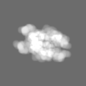
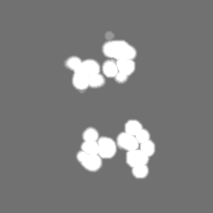
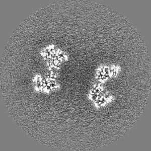
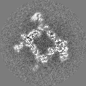
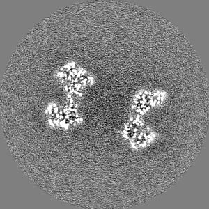
| Projections & Slices |
| ||||||||||||
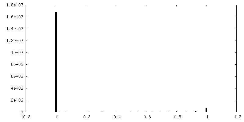
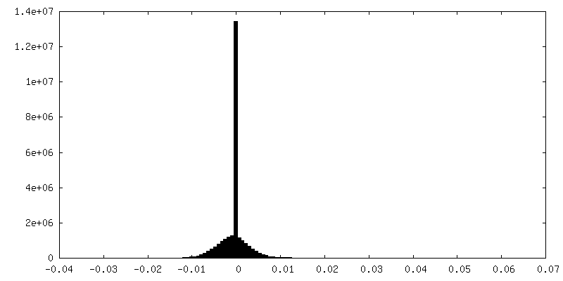
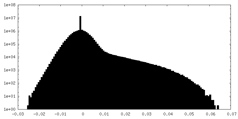
| Density Histograms |
 Z
Z Y
Y X
X


-Half map: #2
| File | emd_30946_half_map_1.map | ||||||||||||
|---|---|---|---|---|---|---|---|---|---|---|---|---|---|
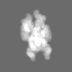
| Projections & Slices |
| ||||||||||||
| Density Histograms |


-Half map: #1
| File | emd_30946_half_map_2.map | ||||||||||||
|---|---|---|---|---|---|---|---|---|---|---|---|---|---|
| Projections & Slices |
| ||||||||||||
| Density Histograms |


- Sample components
-Entire : The octamer of glycoside hydrolase family 57.
| Entire | Name: The octamer of glycoside hydrolase family 57. |
|---|---|
| Components |
|
-Supramolecule #1: The octamer of glycoside hydrolase family 57.
| Supramolecule | Name: The octamer of glycoside hydrolase family 57. / type: complex / ID: 1 / Parent: 0 / Macromolecule list: all |
|---|---|
| Source (natural) | Organism: Staphylothermus marinus (archaea) |
| Recombinant expression | Organism:  Escherichia coli (E. coli) Escherichia coli (E. coli) |
-Macromolecule #1: Glycoside hydrolase, family 57
| Macromolecule | Name: Glycoside hydrolase, family 57 / type: protein_or_peptide / ID: 1 / Number of copies: 8 / Enantiomer: LEVO |
|---|---|
| Source (natural) | Organism: Staphylothermus marinus (strain ATCC 43588 / DSM 3639 / JCM 9404 / F1) (archaea) Strain: ATCC 43588 / DSM 3639 / JCM 9404 / F1 |
| Molecular weight | Theoretical: 75.983641 KDa |
| Recombinant expression | Organism: Escherichia coli BL21(DE3) (bacteria) |
| Sequence | String: LEVLDKYSSL IKPKLINNIE AYMVFDKPAH KPNAEAKIYV LLNNHGSRRD IHYKIVSIDR NREVFSKRIN VDEKKFLIET ISIETPDKP GRFCYKLFID NEQIDNTCFL VGDPSSREQM YFTIVWHHHQ APNYLPDGRI HGPWAYIYVW SDLLKPYGKG P YHYHSVML ...String: LEVLDKYSSL IKPKLINNIE AYMVFDKPAH KPNAEAKIYV LLNNHGSRRD IHYKIVSIDR NREVFSKRIN VDEKKFLIET ISIETPDKP GRFCYKLFID NEQIDNTCFL VGDPSSREQM YFTIVWHHHQ APNYLPDGRI HGPWAYIYVW SDLLKPYGKG P YHYHSVML NIHPHFKATY NLSPSLLRQW QIAVEKGVEF VNGEKYDPNH EKIRLVEETL NNYREALFKG QIDVLTSIYA HT IGGFLTD VLGATNIVEE EIRYGKEVTS KIMGNNYNPQ GIWTPEMAFS MKLIPIYYDL DIKYTVLDDK FHFFHAEGNK DSQ YEPYMV IDTESKKYIT VFFRDHDLSD ILGFRNNFYS EPHAWRNAYE FALRVAEKWF DKNVKVLTIA LDGENWMSFS VNPP LTAYF LDKMIIYLET LSDNKFIKLS TLREIYNKVP ANRILTNIPT NSWLGTFRKW RGEVPQHEEY WIKTYSVYRK LLAYE EMIG GRDEFSNEAR WALWHALDSD YWWAEFWLPK IIDTWLSVAE NILNNRINKI QIIDVRPASE FYEDEKAGLV VTIRNQ LEK EIRVSFAIGG TGFSSVNNDL ETVKMNPNSS YTRIIPVKAK FIGKHKMVVS AISKGLIIDS KIIDINVKPK LLPNPRL EH HHHHH |
-Experimental details
-Structure determination
| Method | negative staining, cryo EM |
|---|---|
 Processing Processing | single particle reconstruction |
| Aggregation state | particle |
-Sample preparation
| Concentration | 2.0 mg/mL |
|---|---|
| Buffer | pH: 7.5 |
| Staining | Type: NEGATIVE / Material: Uranyl acetate |
| Grid | Material: COPPER |
| Vitrification | Cryogen name: ETHANE / Chamber humidity: 100 % / Chamber temperature: 277 K / Instrument: FEI VITROBOT MARK I |
- Electron microscopy
| Microscope | FEI TITAN KRIOS |
|---|---|
| Electron beam | Acceleration voltage: 300 kV / Electron source: FIELD EMISSION GUN |
| Electron optics | Illumination mode: FLOOD BEAM / Imaging mode: BRIGHT FIELDBright-field microscopy / Cs: 2.7 mm |
| Image recording | Film or detector model: GATAN K2 SUMMIT (4k x 4k) / Detector mode: COUNTING / Average exposure time: 8.0 sec. / Average electron dose: 64.0 e/Å2 |
| Experimental equipment |  Model: Titan Krios / Image courtesy: FEI Company |
-Image processing
| Startup model | Type of model: OTHER / Details: Ab initio model from Relion |
|---|---|
| Initial angle assignment | Type: PROJECTION MATCHING |
| Final angle assignment | Type: MAXIMUM LIKELIHOOD |
| Final reconstruction | Resolution.type: BY AUTHOR / Resolution: 2.9 Å / Resolution method: FSC 0.143 CUT-OFF / Number images used: 272770 |
| FSC plot (resolution estimation) | 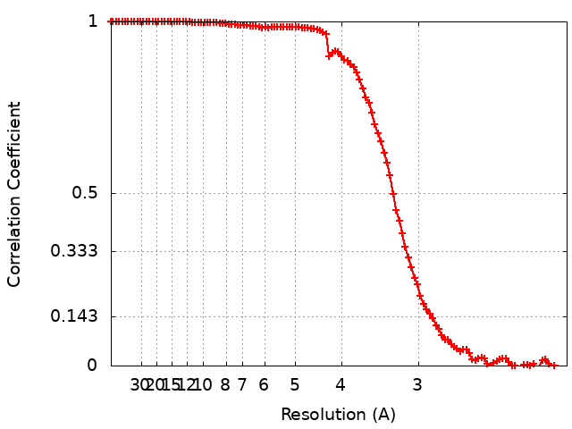 |
-Atomic model buiding 1
| Refinement | Space: REAL / Protocol: AB INITIO MODEL |
|---|---|
| Output model | PDB-7e1y: |
