 Movie
Movie Controller

[English] 日本語
 Yorodumi
- PDB-7d89: Crystal structure of an inactivated double mutant (E182AE280A) of... -
+ Open data

- Basic information
| Entry | Database: PDB / ID: 7d89 | ||||||
|---|---|---|---|---|---|---|---|
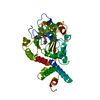
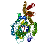
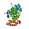
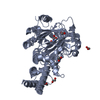
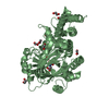
| Title | Crystal structure of an inactivated double mutant (E182AE280A) of a novel thermostable GH10 xylanase XynA | ||||||
 Components Components | Beta-xylanase Xylanase Xylanase | ||||||
 Keywords Keywords | HYDROLASE / GH10 family / Thermostable / Inactivated / mutant | ||||||
| Function / homology |  Function and homology informationendo-1,4-beta-xylanase activity / endo-1,4-beta-xylanase / xylan catabolic process Function and homology informationendo-1,4-beta-xylanase activity / endo-1,4-beta-xylanase / xylan catabolic processSimilarity search - Function | ||||||
| Biological species |  | ||||||
| Method | X-RAY DIFFRACTION / SYNCHROTRON / MOLECULAR REPLACEMENT / Resolution: 2.89384086853 Å | ||||||
 Authors Authors | Xie, W. / Yu, Q. / Wang, C. | ||||||
 Citation Citation | Journal: Biochemistry / Year: 2021 Title: Insights into the Catalytic Mechanism of a Novel XynA and Structure-Based Engineering for Improving Bifunctional Activities. Authors: Xie, W. / Yu, Q. / Zhang, R. / Liu, Y. / Cao, R. / Wang, S. / Zhan, R. / Liu, Z. / Wang, K. / Wang, C. | ||||||
| History |
|
- Structure visualization
| Structure viewer | Molecule: MolmilJmol/JSmol |
|---|
- Downloads & links
-Download
| PDBx/mmCIF format | 7d89.cif.gz | 105 KB | Display | PDBx/mmCIF format |
|---|---|---|---|---|
| PDB format | pdb7d89.ent.gz | 63.1 KB | Display | PDB format |
| PDBx/mmJSON format | 7d89.json.gz | Tree view | PDBx/mmJSON format | |
| Others |  Other downloads Other downloads |
-Validation report
| Arichive directory | https://data.pdbj.org/pub/pdb/validation_reports/d8/7d89ftp://data.pdbj.org/pub/pdb/validation_reports/d8/7d89 | HTTPS FTP |
|---|
-Related structure data
| Related structure data | 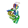 7d88C  1v0lS S: Starting model for refinement C: citing same article ( |
|---|---|
| Similar structure data |


-Links
 PDBj
PDBj
- Assembly
| Deposited unit | 
| ||||||||||||
|---|---|---|---|---|---|---|---|---|---|---|---|---|---|
| 1 |
| ||||||||||||
| Unit cell |
|
-Components
| #1: Protein | Xylanase Mass: 49806.918 Da / Num. of mol.: 1 / Mutation: E182A, E280A Source method: isolated from a genetically manipulated source Source: (gene. exp.) Escherichia coli BL21(DE3) (bacteria) / References: UniProt: A0A4P8ESF9, endo-1,4-beta-xylanase | ||||
|---|---|---|---|---|---|
| #2: Chemical |   Mass: 40.078 Da / Num. of mol.: 2 / Source method: obtained synthetically / Formula: Ca Mass: 40.078 Da / Num. of mol.: 2 / Source method: obtained synthetically / Formula: Ca#3: Water | ChemComp-HOH / | Water Mass: 18.015 Da / Num. of mol.: 2 / Source method: isolated from a natural source / Formula: H2O Mass: 18.015 Da / Num. of mol.: 2 / Source method: isolated from a natural source / Formula: H2OHas ligand of interest | N | |
-Experimental details
-Experiment
| Experiment | Method: X-RAY DIFFRACTION / Number of used crystals: 1 |
|---|
- Sample preparation
| Crystal | Density Matthews: 2.59 Å3/Da / Density % sol: 52.55 % |
|---|---|
| Crystal grow | Temperature: 298 K / Method: vapor diffusion / pH: 6 / Details: 24% PEG 600, 0.1M CaCl2, 0.1M MES pH 6.0 |
-Data collection
| Diffraction | Mean temperature: 100 K / Serial crystal experiment: N |
|---|---|
| Diffraction source | Source: SYNCHROTRON / Site: SSRF  / Beamline: BL19U1 / Wavelength: 0.979 Å / Beamline: BL19U1 / Wavelength: 0.979 Å |
| Detector | Type: ADSC QUANTUM 315r / Detector: CCD / Date: Jul 8, 2019 |
| Radiation | Protocol: SINGLE WAVELENGTH / Monochromatic (M) / Laue (L): M / Scattering type: x-ray |
| Radiation wavelength | Wavelength: 0.979 Å / Relative weight: 1 |
| Reflection | Resolution: 2.89→49.28 Å / Num. obs: 12123 / % possible obs: 99.56 % / Redundancy: 25.5 % / Biso Wilson estimate: 66.2367949314 Å2 / CC1/2: 1 / Rmerge(I) obs: 0.108 / Rpim(I) all: 0.021 / Rrim(I) all: 0.108 / Net I/σ(I): 34 |
| Reflection shell | Resolution: 2.894→2.997 Å / Rmerge(I) obs: 0.108 / Num. unique obs: 12123 / CC1/2: 1 / Rpim(I) all: 0.021 / Rrim(I) all: 0.108 |
- Processing
| Software |
| |||||||||||||||||||||||||||||||||||
|---|---|---|---|---|---|---|---|---|---|---|---|---|---|---|---|---|---|---|---|---|---|---|---|---|---|---|---|---|---|---|---|---|---|---|---|---|
| Refinement | Method to determine structure: MOLECULAR REPLACEMENT Starting model: 1V0L Resolution: 2.89384086853→49.2798077289 Å / SU ML: 0.397124008712 / Cross valid method: FREE R-VALUE / σ(F): 1.37753762711 / Phase error: 28.1305524779 Stereochemistry target values: GeoStd + Monomer Library + CDL v1.2
| |||||||||||||||||||||||||||||||||||
| Solvent computation | Shrinkage radii: 0.9 Å / VDW probe radii: 1.11 Å / Solvent model: FLAT BULK SOLVENT MODEL | |||||||||||||||||||||||||||||||||||
| Displacement parameters | Biso mean: 58.640519927 Å2 | |||||||||||||||||||||||||||||||||||
| Refinement step | Cycle: LAST / Resolution: 2.89384086853→49.2798077289 Å
| |||||||||||||||||||||||||||||||||||
| Refine LS restraints |
| |||||||||||||||||||||||||||||||||||
| LS refinement shell |
|
