 Movie
Movie Controller


+ Open data

- Basic information
| Entry | Database: PDB / ID: 1jxo | ||||||
|---|---|---|---|---|---|---|---|
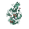
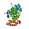
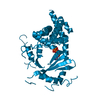
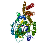
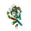
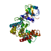
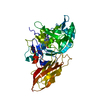
| Title | Crystal Structure of the SH3-HOOK-GK Fragment of PSD-95 | ||||||
 Components Components | postsynaptic density protein | ||||||
 Keywords Keywords | STRUCTURAL PROTEIN / MAGUK / postsynaptic density / SH3 domain / guanylate kinase domain | ||||||
| Function / homology |  Function and homology information Function and homology informationRHO GTPases activate CIT / positive regulation of AMPA glutamate receptor clustering / neuronal ion channel clustering / P2Y1 nucleotide receptor binding / beta-1 adrenergic receptor binding / Neurexins and neuroligins / neuroligin family protein binding / receptor localization to synapse / positive regulation of neuron projection arborization / regulation of grooming behavior ...RHO GTPases activate CIT / positive regulation of AMPA glutamate receptor clustering / neuronal ion channel clustering / P2Y1 nucleotide receptor binding / beta-1 adrenergic receptor binding / Neurexins and neuroligins / neuroligin family protein binding / receptor localization to synapse / positive regulation of neuron projection arborization / regulation of grooming behavior / structural constituent of postsynaptic density / synaptic vesicle maturation / proximal dendrite / AMPA glutamate receptor clustering / cerebellar mossy fiber / protein localization to synapse / cellular response to potassium ion / vocalization behavior / LGI-ADAM interactions / neuron spine / Trafficking of AMPA receptors / dendritic branch / Activation of Ca-permeable Kainate Receptor / juxtaparanode region of axon / neuron projection terminus / establishment or maintenance of epithelial cell apical/basal polarity / dendritic spine morphogenesis / negative regulation of receptor internalization / postsynaptic neurotransmitter receptor diffusion trapping / frizzled binding / dendritic spine organization / acetylcholine receptor binding / positive regulation of synapse assembly / RAF/MAP kinase cascade / Synaptic adhesion-like molecules / neurotransmitter receptor localization to postsynaptic specialization membrane / positive regulation of dendrite morphogenesis / beta-2 adrenergic receptor binding / regulation of neuronal synaptic plasticity / locomotory exploration behavior / cortical cytoskeleton / regulation of NMDA receptor activity / social behavior / positive regulation of excitatory postsynaptic potential / AMPA glutamate receptor complex / kinesin binding / neuromuscular process controlling balance / excitatory synapse / D1 dopamine receptor binding / Unblocking of NMDA receptors, glutamate binding and activation / glutamate receptor binding / positive regulation of protein tyrosine kinase activity / positive regulation of synaptic transmission / ionotropic glutamate receptor binding / extrinsic component of cytoplasmic side of plasma membrane / dendrite cytoplasm / synaptic membrane / PDZ domain binding / cell periphery / postsynaptic density membrane / regulation of long-term neuronal synaptic plasticity / neuromuscular junction / establishment of protein localization / cell-cell adhesion / cerebral cortex development / kinase binding / cell-cell junction / synaptic vesicle / cell junction / positive regulation of cytosolic calcium ion concentration / chemical synaptic transmission / postsynaptic membrane / postsynapse / scaffold protein binding / basolateral plasma membrane / protein-containing complex assembly / protein phosphatase binding / dendritic spine / postsynaptic density / neuron projection / signaling receptor binding / dendrite / glutamatergic synapse / synapse / protein-containing complex binding / protein kinase binding / endoplasmic reticulum / membrane / plasma membrane / cytosol / cytoplasmSimilarity search - Function | ||||||
| Biological species |  Rattus norvegicus (Norway rat) Rattus norvegicus (Norway rat) | ||||||
| Method | X-RAY DIFFRACTION / SYNCHROTRON / MOLECULAR REPLACEMENT / Resolution: 2.3 Å | ||||||
 Authors Authors | Tavares, G.A. / Panepucci, E.H. / Brunger, A.T. | ||||||
 Citation Citation | Journal: Mol.Cell / Year: 2001 Title: Structural characterization of the intramolecular interaction between the SH3 and guanylate kinase domains of PSD-95. Authors: Tavares, G.A. / Panepucci, E.H. / Brunger, A.T. | ||||||
| History |
|
- Structure visualization
| Structure viewer | Molecule: MolmilJmol/JSmol |
|---|
- Downloads & links
-Download
| PDBx/mmCIF format | 1jxo.cif.gz | 117.2 KB | Display | PDBx/mmCIF format |
|---|---|---|---|---|
| PDB format | pdb1jxo.ent.gz | 87.9 KB | Display | PDB format |
| PDBx/mmJSON format | 1jxo.json.gz | Tree view | PDBx/mmJSON format | |
| Others |  Other downloads Other downloads |
-Validation report
| Arichive directory | https://data.pdbj.org/pub/pdb/validation_reports/jx/1jxoftp://data.pdbj.org/pub/pdb/validation_reports/jx/1jxo | HTTPS FTP |
|---|
-Related structure data
| Related structure data | 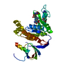 1jxmSC S: Starting model for refinement C: citing same article ( |
|---|---|
| Similar structure data |


-Links
 PDBj
PDBj


- Assembly
| Deposited unit | 
| ||||||||
|---|---|---|---|---|---|---|---|---|---|
| 1 | 
| ||||||||
| 2 | 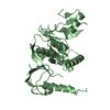
| ||||||||
| Unit cell |
|
-Components
| #1: Protein | / POSTSYNAPTIC DENSITY-95 / PSD-95 Mass: 34812.996 Da / Num. of mol.: 2 / Fragment: SH3-HOOK-GK Source method: isolated from a genetically manipulated source Source: (gene. exp.) Rattus norvegicus (Norway rat) / Gene: PSD-95 / Organ: brain / Plasmid: pET28a / Production host:  Escherichia coli (E. coli) / Strain (production host): BL21DE3* / References: UniProt: P31016 Escherichia coli (E. coli) / Strain (production host): BL21DE3* / References: UniProt: P31016#2: Water | ChemComp-HOH / | Water Mass: 18.015 Da / Num. of mol.: 188 / Source method: isolated from a natural source / Formula: H2O Mass: 18.015 Da / Num. of mol.: 188 / Source method: isolated from a natural source / Formula: H2O |
|---|
-Experimental details
-Experiment
| Experiment | Method: X-RAY DIFFRACTION / Number of used crystals: 1 |
|---|
- Sample preparation
| Crystal | Density Matthews: 2.18 Å3/Da / Density % sol: 43.64 % | ||||||||||||||||||||||||||||||
|---|---|---|---|---|---|---|---|---|---|---|---|---|---|---|---|---|---|---|---|---|---|---|---|---|---|---|---|---|---|---|---|
| Crystal grow | Temperature: 277 K / Method: vapor diffusion, hanging drop / pH: 6.3 Details: 2-methyl-2,4-pentanediol, MES buffer, pH 6.3, VAPOR DIFFUSION, HANGING DROP, temperature 277K | ||||||||||||||||||||||||||||||
| Crystal grow | *PLUS Temperature: 4 ℃ | ||||||||||||||||||||||||||||||
| Components of the solutions | *PLUS
|
-Data collection
| Diffraction | Mean temperature: 100 K |
|---|---|
| Diffraction source | Source: SYNCHROTRON / Site: SSRL  / Beamline: BL11-1 / Wavelength: 0.964818 Å / Beamline: BL11-1 / Wavelength: 0.964818 Å |
| Detector | Type: ADSC QUANTUM 4 / Detector: CCD / Date: May 19, 2001 Details: Flat mirror (vertical focusing); single crystal Si(111) bent monochromator (horizontal focusing) |
| Radiation | Monochromator: Flat mirror (vertical focusing); single crystal Si(111) bent monochromator (horizontal focusing) Protocol: SINGLE WAVELENGTH / Monochromatic (M) / Laue (L): M / Scattering type: x-ray |
| Radiation wavelength | Wavelength: 0.964818 Å / Relative weight: 1 |
| Reflection | Resolution: 2.3→29.88 Å / Num. all: 25724 / Num. obs: 25456 / % possible obs: 97.3 % / Observed criterion σ(F): 0 / Redundancy: 3 % / Biso Wilson estimate: 20 Å2 / Rsym value: 0.043 |
| Reflection shell | Resolution: 2.3→2.34 Å / Redundancy: 3 % / Num. unique all: 1270 / Rsym value: 0.106 / % possible all: 97.6 |
| Reflection | *PLUS Num. obs: 25724 / % possible obs: 98.4 % / Rmerge(I) obs: 0.043 |
| Reflection shell | *PLUS % possible obs: 97.6 % / Rmerge(I) obs: 0.102 |
- Processing
| Software |
| ||||||||||||||||||||||||||||||||||||||||||||||||||||||||||||||||||||||||||||||||
|---|---|---|---|---|---|---|---|---|---|---|---|---|---|---|---|---|---|---|---|---|---|---|---|---|---|---|---|---|---|---|---|---|---|---|---|---|---|---|---|---|---|---|---|---|---|---|---|---|---|---|---|---|---|---|---|---|---|---|---|---|---|---|---|---|---|---|---|---|---|---|---|---|---|---|---|---|---|---|---|---|---|
| Refinement | Method to determine structure: MOLECULAR REPLACEMENT Starting model: PDB ENTRY 1JXM Resolution: 2.3→29.88 Å / Rfactor Rfree error: 0.005 / Data cutoff high absF: 1763834.74 / Data cutoff low absF: 0 / Isotropic thermal model: RESTRAINED / Cross valid method: THROUGHOUT / σ(F): 0 / Stereochemistry target values: Engh & Huber
| ||||||||||||||||||||||||||||||||||||||||||||||||||||||||||||||||||||||||||||||||
| Solvent computation | Solvent model: FLAT MODEL / Bsol: 38.191 Å2 / ksol: 0.335299 e/Å3 | ||||||||||||||||||||||||||||||||||||||||||||||||||||||||||||||||||||||||||||||||
| Displacement parameters | Biso mean: 28.6 Å2
| ||||||||||||||||||||||||||||||||||||||||||||||||||||||||||||||||||||||||||||||||
| Refine analyze |
| ||||||||||||||||||||||||||||||||||||||||||||||||||||||||||||||||||||||||||||||||
| Refinement step | Cycle: LAST / Resolution: 2.3→29.88 Å
| ||||||||||||||||||||||||||||||||||||||||||||||||||||||||||||||||||||||||||||||||
| Refine LS restraints |
| ||||||||||||||||||||||||||||||||||||||||||||||||||||||||||||||||||||||||||||||||
| LS refinement shell | Resolution: 2.3→2.38 Å / Rfactor Rfree error: 0.02 / Total num. of bins used: 10
| ||||||||||||||||||||||||||||||||||||||||||||||||||||||||||||||||||||||||||||||||
| Xplor file |
| ||||||||||||||||||||||||||||||||||||||||||||||||||||||||||||||||||||||||||||||||
| Software | *PLUS Name: CNS / Version: 1.1 / Classification: refinement | ||||||||||||||||||||||||||||||||||||||||||||||||||||||||||||||||||||||||||||||||
| Refinement | *PLUS % reflection Rfree: 9.9 % / Rfactor obs: 0.22 / Rfactor Rwork: 0.22 | ||||||||||||||||||||||||||||||||||||||||||||||||||||||||||||||||||||||||||||||||
| Solvent computation | *PLUS | ||||||||||||||||||||||||||||||||||||||||||||||||||||||||||||||||||||||||||||||||
| Displacement parameters | *PLUS Biso mean: 28.6 Å2 | ||||||||||||||||||||||||||||||||||||||||||||||||||||||||||||||||||||||||||||||||
| Refine LS restraints | *PLUS
| ||||||||||||||||||||||||||||||||||||||||||||||||||||||||||||||||||||||||||||||||
| LS refinement shell | *PLUS Rfactor Rfree: 0.298 / % reflection Rfree: 8.9 % / Rfactor Rwork: 0.222 |
