Resolution: 1.56→36.97 Å / Cor.coef. Fo:Fc: 0.96 / Cor.coef. Fo:Fc free: 0.935 / SU B: 5.985 / SU ML: 0.098 / Cross valid method: THROUGHOUT / ESU R: 0.112 / ESU R Free: 0.102 / Stereochemistry target values: MAXIMUM LIKELIHOOD / Details: HYDROGENS HAVE BEEN ADDED IN THE RIDING POSITIONS
Rfactor
Num. reflection
% reflection
Selection details
Rfree
0.25844
3667
5 %
RANDOM
Rwork
0.198
-
-
-
obs
0.20108
69608
94.19 %
-
Solvent computation
Ion probe radii: 0.8 Å / Shrinkage radii: 0.8 Å / VDW probe radii: 1.2 Å / Solvent model: MASK
 Movie
Movie Controller

 Yorodumi
 Open data
 Basic information
 Components
 Keywords
 HYDROLASE /
  Function and homology information
 Authors
 Japan, 1items
  Citation
 Structure visualization
 Downloads & links
 Other downloads


 PDBj
PDBj


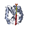
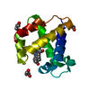
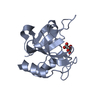
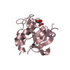
 Assembly
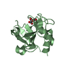


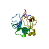
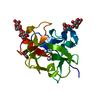
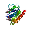
 Mass: 189.100 Da / Num. of mol.: 4 / Source method: obtained synthetically / Formula: C6H5O7 / Feature type: SUBJECT OF INVESTIGATION
 Mass: 18.015 Da / Num. of mol.: 246 / Source method: isolated from a natural source / Formula: H2O
 Sample preparation
 Processing