 Movie
Movie Controller

[English] 日本語
 Yorodumi
- PDB-1knm: Streptomyces lividans Xylan Binding Domain cbm13 in Complex with ... -
+ Open data

- Basic information
| Entry | Database: PDB / ID: 1knm | |||||||||
|---|---|---|---|---|---|---|---|---|---|---|
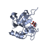
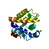
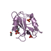
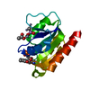
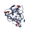
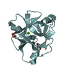
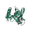
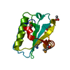
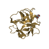
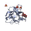
| Title | Streptomyces lividans Xylan Binding Domain cbm13 in Complex with Lactose | |||||||||
 Components Components | ENDO-1,4-BETA-XYLANASE A | |||||||||
 Keywords Keywords |  HYDROLASE / xylanase A xylan binding domain cbm13 / lectin-like beta trefoil fold / lactose complex HYDROLASE / xylanase A xylan binding domain cbm13 / lectin-like beta trefoil fold / lactose complex | |||||||||
| Function / homology |  Function and homology informationendo-1,4-beta-xylanase activity / endo-1,4-beta-xylanase / xylan catabolic process / carbohydrate binding / extracellular region Function and homology informationendo-1,4-beta-xylanase activity / endo-1,4-beta-xylanase / xylan catabolic process / carbohydrate binding / extracellular regionSimilarity search - Function | |||||||||
| Biological species |  Streptomyces lividans (bacteria) Streptomyces lividans (bacteria) | |||||||||
| Method | X-RAY DIFFRACTION / SYNCHROTRON / MOLECULAR REPLACEMENT / Resolution: 1.2 Å | |||||||||
 Authors Authors | Notenboom, V. / Boraston, A.B. / Williams, S.J. / Kilburn, D.G. / Rose, D.R. | |||||||||
 Citation Citation | Journal: Biochemistry / Year: 2002 Title: High-resolution crystal structures of the lectin-like xylan binding domain from Streptomyces lividans xylanase 10A with bound substrates reveal a novel mode of xylan binding. Authors: Notenboom, V. / Boraston, A.B. / Williams, S.J. / Kilburn, D.G. / Rose, D.R. | |||||||||
| History |
|
- Structure visualization
| Structure viewer | Molecule: MolmilJmol/JSmol |
|---|
- Downloads & links
-Download
| PDBx/mmCIF format | 1knm.cif.gz | 40.9 KB | Display | PDBx/mmCIF format |
|---|---|---|---|---|
| PDB format | pdb1knm.ent.gz | 30 KB | Display | PDB format |
| PDBx/mmJSON format | 1knm.json.gz | Tree view | PDBx/mmJSON format | |
| Others |  Other downloads Other downloads |
-Validation report
| Arichive directory | https://data.pdbj.org/pub/pdb/validation_reports/kn/1knmftp://data.pdbj.org/pub/pdb/validation_reports/kn/1knm | HTTPS FTP |
|---|
-Related structure data


-Links
 PDBj
PDBj


- Assembly
| Deposited unit | 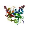
| ||||||||
|---|---|---|---|---|---|---|---|---|---|
| 1 |
| ||||||||
| Unit cell |
|
-Components
| #1: Protein | Mass: 13608.731 Da / Num. of mol.: 1 / Fragment: carbohydrate binding module (residues 348-477) Source method: isolated from a genetically manipulated source Source: (gene. exp.) Streptomyces lividans (bacteria) / Production host: Escherichia coli (E. coli) / References: UniProt: P26514, endo-1,4-beta-xylanase | ||||
|---|---|---|---|---|---|
| #2: Polysaccharide |   , Oligosaccharide / Class: Nutrient / Mass: 342.297 Da / Num. of mol.: 2 , Oligosaccharide / Class: Nutrient / Mass: 342.297 Da / Num. of mol.: 2Source method: isolated from a genetically manipulated source Details: oligosaccharide / References: beta-lactose #3: Chemical | ChemComp-GOL / | Glycerol  Mass: 92.094 Da / Num. of mol.: 1 / Source method: obtained synthetically / Formula: C3H8O3 Mass: 92.094 Da / Num. of mol.: 1 / Source method: obtained synthetically / Formula: C3H8O3#4: Water | ChemComp-HOH / | Water Mass: 18.015 Da / Num. of mol.: 237 / Source method: isolated from a natural source / Formula: H2O Mass: 18.015 Da / Num. of mol.: 237 / Source method: isolated from a natural source / Formula: H2O |
-Experimental details
-Experiment
| Experiment | Method: X-RAY DIFFRACTION / Number of used crystals: 1 |
|---|
- Sample preparation
| Crystal | Density Matthews: 2.07 Å3/Da / Density % sol: 40.56 % | ||||||||||||||||||||||||||||||
|---|---|---|---|---|---|---|---|---|---|---|---|---|---|---|---|---|---|---|---|---|---|---|---|---|---|---|---|---|---|---|---|
| Crystal grow | Temperature: 300 K / Method: vapor diffusion, hanging drop / pH: 6.5 Details: 1.8 mM Ammonium Sulfate, 50 mM MES pH 6.5, 5% dioxane, VAPOR DIFFUSION, HANGING DROP, temperature 300K | ||||||||||||||||||||||||||||||
| Crystal grow | *PLUS Method: vapor diffusion | ||||||||||||||||||||||||||||||
| Components of the solutions | *PLUS
|
-Data collection
| Diffraction | Mean temperature: 100 K |
|---|---|
| Diffraction source | Source: SYNCHROTRON / Site: APS  / Beamline: 14-BM-D / Wavelength: 1 Å / Beamline: 14-BM-D / Wavelength: 1 Å |
| Detector | Type: ADSC QUANTUM 4 / Detector: CCD / Date: Mar 19, 2000 |
| Radiation | Monochromator: graphite / Protocol: SINGLE WAVELENGTH / Monochromatic (M) / Laue (L): M / Scattering type: x-ray |
| Radiation wavelength | Wavelength: 1 Å / Relative weight: 1 |
| Reflection | Resolution: 1.2→1.24 Å / Num. obs: 30016 / % possible obs: 83.1 % / Observed criterion σ(F): 2 / Observed criterion σ(I): 2 |
| Reflection shell | Highest resolution: 1.2 Å / % possible all: 83.1 |
| Reflection | *PLUS Num. all: 126516 / Rmerge(I) obs: 0.041 |
| Reflection shell | *PLUS % possible obs: 22.1 % / Rmerge(I) obs: 0.193 / Mean I/σ(I) obs: 9.3 |
- Processing
| Software |
| |||||||||||||||||||||||||
|---|---|---|---|---|---|---|---|---|---|---|---|---|---|---|---|---|---|---|---|---|---|---|---|---|---|---|
| Refinement | Method to determine structure: MOLECULAR REPLACEMENT / Resolution: 1.2→19.57 Å / σ(F): 0 / Stereochemistry target values: refmac5
| |||||||||||||||||||||||||
| Refinement step | Cycle: LAST / Resolution: 1.2→19.57 Å
| |||||||||||||||||||||||||
| Refine LS restraints |
| |||||||||||||||||||||||||
| LS refinement shell | Resolution: 1.2→1.24 Å /
| |||||||||||||||||||||||||
| Refinement | *PLUS % reflection Rfree: 5 % | |||||||||||||||||||||||||
| Solvent computation | *PLUS | |||||||||||||||||||||||||
| Displacement parameters | *PLUS | |||||||||||||||||||||||||
| Refine LS restraints | *PLUS
| |||||||||||||||||||||||||
| LS refinement shell | *PLUS Rfactor Rwork: 0.23 |
