 Movie
Movie Controller

[English] 日本語
 Yorodumi
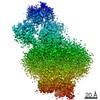
- PDB-7ceg: Crystal structure of the complex between mouse PTP delta and neur... -
+ Open data

- Basic information
| Entry | Database: PDB / ID: 7ceg | |||||||||||||||
|---|---|---|---|---|---|---|---|---|---|---|---|---|---|---|---|---|
| Title | Crystal structure of the complex between mouse PTP delta and neuroligin-3 | |||||||||||||||
 Components Components |
| |||||||||||||||
 Keywords Keywords | HYDROLASE/CELL ADHESION / synapse organization / trans-synaptic complex /  esterase domain / HYDROLASE-CELL ADHESION complex esterase domain / HYDROLASE-CELL ADHESION complex | |||||||||||||||
| Function / homology |  Function and homology information Function and homology informationrhythmic synaptic transmission / Neurexins and neuroligins / Receptor-type tyrosine-protein phosphatases / trans-synaptic signaling / Synaptic adhesion-like molecules / trans-synaptic signaling by trans-synaptic complex / postsynaptic specialization assembly / regulation of terminal button organization / negative regulation of dendritic spine morphogenesis / postsynaptic membrane assembly ...rhythmic synaptic transmission / Neurexins and neuroligins / Receptor-type tyrosine-protein phosphatases / trans-synaptic signaling / Synaptic adhesion-like molecules / trans-synaptic signaling by trans-synaptic complex / postsynaptic specialization assembly / regulation of terminal button organization / negative regulation of dendritic spine morphogenesis / postsynaptic membrane assembly / positive regulation of synaptic vesicle clustering / circadian sleep/wake cycle / presynaptic membrane assembly / synaptic membrane adhesion / regulation of postsynaptic density assembly / inhibitory synapse / regulation of respiratory gaseous exchange by nervous system process / neuron cell-cell adhesion / neurexin family protein binding / negative regulation of excitatory postsynaptic potential / vocalization behavior / negative regulation of receptor signaling pathway via JAK-STAT / regulation of AMPA receptor activity / regulation of dendritic spine morphogenesis / positive regulation of inhibitory postsynaptic potential / postsynaptic specialization membrane / inhibitory postsynaptic potential / axon extension / positive regulation of synapse assembly / regulation of long-term synaptic potentiation / positive regulation of AMPA receptor activity / positive regulation of dendrite morphogenesis / regulation of NMDA receptor activity / positive regulation of protein localization to synapse / heterophilic cell-cell adhesion via plasma membrane cell adhesion molecules / adult behavior / positive regulation of dendritic spine development / oligodendrocyte differentiation / social behavior / positive regulation of excitatory postsynaptic potential / synaptic vesicle endocytosis / excitatory synapse / endocytic vesicle / regulation of immune response / GABA-ergic synapse / prepulse inhibition / dephosphorylation / regulation of synaptic transmission, glutamatergic / cell adhesion molecule binding / synapse assembly / excitatory postsynaptic potential / hippocampal mossy fiber to CA3 synapse / receptor-mediated endocytosis / positive regulation of synaptic transmission, glutamatergic / protein-tyrosine-phosphatase / learning / protein tyrosine phosphatase activity / long-term synaptic potentiation / synapse organization / Schaffer collateral - CA1 synapse / visual learning / modulation of chemical synaptic transmission / neuron differentiation / presynapse / presynaptic membrane / signaling receptor activity / scaffold protein binding / chemical synaptic transmission / postsynaptic membrane / molecular adaptor activity / receptor complex / signaling receptor binding / neuronal cell body / glutamatergic synapse / synapse / dendrite / cell surface / plasma membraneSimilarity search - Function | |||||||||||||||
| Biological species |  Mus musculus (house mouse) Mus musculus (house mouse) | |||||||||||||||
| Method | X-RAY DIFFRACTION / SYNCHROTRON / MOLECULAR REPLACEMENT / Resolution: 3.85 Å | |||||||||||||||
 Authors Authors | Yamagata, A. / Yoshida, T. / Shiroshima, T. / Maeda, A. / Fukai, S. | |||||||||||||||
| Funding support |  Japan, 4items Japan, 4items
| |||||||||||||||
 Citation Citation | Journal: Nat Commun / Year: 2021 Title: Canonical versus non-canonical transsynaptic signaling of neuroligin 3 tunes development of sociality in mice. Authors: Yoshida, T. / Yamagata, A. / Imai, A. / Kim, J. / Izumi, H. / Nakashima, S. / Shiroshima, T. / Maeda, A. / Iwasawa-Okamoto, S. / Azechi, K. / Osaka, F. / Saitoh, T. / Maenaka, K. / Shimada, ...Authors: Yoshida, T. / Yamagata, A. / Imai, A. / Kim, J. / Izumi, H. / Nakashima, S. / Shiroshima, T. / Maeda, A. / Iwasawa-Okamoto, S. / Azechi, K. / Osaka, F. / Saitoh, T. / Maenaka, K. / Shimada, T. / Fukata, Y. / Fukata, M. / Matsumoto, J. / Nishijo, H. / Takao, K. / Tanaka, S. / Okabe, S. / Tabuchi, K. / Uemura, T. / Mishina, M. / Mori, H. / Fukai, S. | |||||||||||||||
| History |
|
- Structure visualization
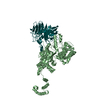
| Structure viewer | Molecule: MolmilJmol/JSmol |
|---|
- Downloads & links
-Download
| PDBx/mmCIF format | 7ceg.cif.gz | 391.9 KB | Display | PDBx/mmCIF format |
|---|---|---|---|---|
| PDB format | pdb7ceg.ent.gz | 320.6 KB | Display | PDB format |
| PDBx/mmJSON format | 7ceg.json.gz | Tree view | PDBx/mmJSON format | |
| Others |  Other downloads Other downloads |
-Validation report
| Arichive directory | https://data.pdbj.org/pub/pdb/validation_reports/ce/7cegftp://data.pdbj.org/pub/pdb/validation_reports/ce/7ceg | HTTPS FTP |
|---|
-Related structure data
| Related structure data |  7ceeC 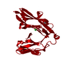 2yd6S  3bixS  4yfdS  4yfeS S: Starting model for refinement C: citing same article ( |
|---|---|
| Similar structure data |


-Links
 PDBj
PDBj


- Assembly
| Deposited unit | 
| ||||||||
|---|---|---|---|---|---|---|---|---|---|
| 1 |
| ||||||||
| Unit cell |
|
-Components
| #1: Protein | Mass: 42384.637 Da / Num. of mol.: 1 Source method: isolated from a genetically manipulated source Source: (gene. exp.) Mus musculus (house mouse) / Gene: Ptprd / Cell line (production host): HEK293F / Production host:  Homo sapiens (human) / References: UniProt: Q64487, protein-tyrosine-phosphatase Homo sapiens (human) / References: UniProt: Q64487, protein-tyrosine-phosphatase | ||
|---|---|---|---|
| #2: Protein | NLGN3 / Gliotactin homolog Mass: 64508.363 Da / Num. of mol.: 1 Source method: isolated from a genetically manipulated source Source: (gene. exp.) Mus musculus (house mouse) / Cell line: HEK293F / Gene: Nlgn3 / Cell line (production host): HEK293F / Production host: Homo sapiens (human) / References: UniProt: Q8BYM5 | ||
| #3: Sugar | ChemComp-NAG / N-Acetylglucosamine  Type: D-saccharide, beta linking / Mass: 221.208 Da / Num. of mol.: 4 Type: D-saccharide, beta linking / Mass: 221.208 Da / Num. of mol.: 4Source method: isolated from a genetically manipulated source Formula: C8H15NO6 Has ligand of interest | N | |
-Experimental details
-Experiment
| Experiment | Method: X-RAY DIFFRACTION / Number of used crystals: 1 |
|---|
- Sample preparation
| Crystal | Density Matthews: 4.28 Å3/Da / Density % sol: 71.28 % |
|---|---|
| Crystal grow | Temperature: 298 K / Method: vapor diffusion, sitting drop / pH: 6 / Details: 10% PEG 3350, 0.1 M ammonium iodide, 0.1 M MES-Na |
-Data collection
| Diffraction | Mean temperature: 100 K / Serial crystal experiment: N | ||||||||||||||||||||||||||||||||||||||||||||||||||||||||||||||||||||||||||||||||||||||||||||||||||||||||||||||||||||||||||||||||||||||||||||||||||||||||||||||||||||||||||||||||||||
|---|---|---|---|---|---|---|---|---|---|---|---|---|---|---|---|---|---|---|---|---|---|---|---|---|---|---|---|---|---|---|---|---|---|---|---|---|---|---|---|---|---|---|---|---|---|---|---|---|---|---|---|---|---|---|---|---|---|---|---|---|---|---|---|---|---|---|---|---|---|---|---|---|---|---|---|---|---|---|---|---|---|---|---|---|---|---|---|---|---|---|---|---|---|---|---|---|---|---|---|---|---|---|---|---|---|---|---|---|---|---|---|---|---|---|---|---|---|---|---|---|---|---|---|---|---|---|---|---|---|---|---|---|---|---|---|---|---|---|---|---|---|---|---|---|---|---|---|---|---|---|---|---|---|---|---|---|---|---|---|---|---|---|---|---|---|---|---|---|---|---|---|---|---|---|---|---|---|---|---|---|---|
| Diffraction source | Source: SYNCHROTRON / Site: SPring-8 / Beamline: BL41XU / Wavelength: 1 Å | ||||||||||||||||||||||||||||||||||||||||||||||||||||||||||||||||||||||||||||||||||||||||||||||||||||||||||||||||||||||||||||||||||||||||||||||||||||||||||||||||||||||||||||||||||||
| Detector | Type: DECTRIS PILATUS3 S 6M / Detector: PIXEL / Date: Nov 18, 2014 | ||||||||||||||||||||||||||||||||||||||||||||||||||||||||||||||||||||||||||||||||||||||||||||||||||||||||||||||||||||||||||||||||||||||||||||||||||||||||||||||||||||||||||||||||||||
| Radiation | Protocol: SINGLE WAVELENGTH / Monochromatic (M) / Laue (L): M / Scattering type: x-ray | ||||||||||||||||||||||||||||||||||||||||||||||||||||||||||||||||||||||||||||||||||||||||||||||||||||||||||||||||||||||||||||||||||||||||||||||||||||||||||||||||||||||||||||||||||||
| Radiation wavelength | Wavelength: 1 Å / Relative weight: 1 | ||||||||||||||||||||||||||||||||||||||||||||||||||||||||||||||||||||||||||||||||||||||||||||||||||||||||||||||||||||||||||||||||||||||||||||||||||||||||||||||||||||||||||||||||||||
| Reflection | Resolution: 3.85→50 Å / Num. obs: 19070 / % possible obs: 98.4 % / Redundancy: 8.9 % / Biso Wilson estimate: 142.02 Å2 / Rmerge(I) obs: 0.119 / Rpim(I) all: 0.034 / Rrim(I) all: 0.124 / Χ2: 0.799 / Net I/σ(I): 4.1 | ||||||||||||||||||||||||||||||||||||||||||||||||||||||||||||||||||||||||||||||||||||||||||||||||||||||||||||||||||||||||||||||||||||||||||||||||||||||||||||||||||||||||||||||||||||
| Reflection shell | Diffraction-ID: 1
|
- Processing
| Software |
| ||||||||||||||||||||||||||||||||||||||||||||||||||||||||||||||||||||||||||||||||||||||||||||||||||||||||||||||||||||||||||||||||||||||||||||||||||||||
|---|---|---|---|---|---|---|---|---|---|---|---|---|---|---|---|---|---|---|---|---|---|---|---|---|---|---|---|---|---|---|---|---|---|---|---|---|---|---|---|---|---|---|---|---|---|---|---|---|---|---|---|---|---|---|---|---|---|---|---|---|---|---|---|---|---|---|---|---|---|---|---|---|---|---|---|---|---|---|---|---|---|---|---|---|---|---|---|---|---|---|---|---|---|---|---|---|---|---|---|---|---|---|---|---|---|---|---|---|---|---|---|---|---|---|---|---|---|---|---|---|---|---|---|---|---|---|---|---|---|---|---|---|---|---|---|---|---|---|---|---|---|---|---|---|---|---|---|---|---|---|---|
| Refinement | Method to determine structure: MOLECULAR REPLACEMENT Starting model: 2YD6, 4YFD, 4YFE, 3BIX Resolution: 3.85→49.69 Å / SU ML: 0.63 / Cross valid method: THROUGHOUT / σ(F): 1.35 / Phase error: 33.37 / Stereochemistry target values: ML
| ||||||||||||||||||||||||||||||||||||||||||||||||||||||||||||||||||||||||||||||||||||||||||||||||||||||||||||||||||||||||||||||||||||||||||||||||||||||
| Solvent computation | Shrinkage radii: 0.9 Å / VDW probe radii: 1.11 Å / Solvent model: FLAT BULK SOLVENT MODEL | ||||||||||||||||||||||||||||||||||||||||||||||||||||||||||||||||||||||||||||||||||||||||||||||||||||||||||||||||||||||||||||||||||||||||||||||||||||||
| Refinement step | Cycle: LAST / Resolution: 3.85→49.69 Å
| ||||||||||||||||||||||||||||||||||||||||||||||||||||||||||||||||||||||||||||||||||||||||||||||||||||||||||||||||||||||||||||||||||||||||||||||||||||||
| Refine LS restraints |
| ||||||||||||||||||||||||||||||||||||||||||||||||||||||||||||||||||||||||||||||||||||||||||||||||||||||||||||||||||||||||||||||||||||||||||||||||||||||
| LS refinement shell |
| ||||||||||||||||||||||||||||||||||||||||||||||||||||||||||||||||||||||||||||||||||||||||||||||||||||||||||||||||||||||||||||||||||||||||||||||||||||||
| Refinement TLS params. | Method: refined / Refine-ID: X-RAY DIFFRACTION
| ||||||||||||||||||||||||||||||||||||||||||||||||||||||||||||||||||||||||||||||||||||||||||||||||||||||||||||||||||||||||||||||||||||||||||||||||||||||
| Refinement TLS group |
|
