 Movie
Movie Controller

[English] 日本語
 Yorodumi
- PDB-7c8h: Ambient temperature structure of Bifidobacterium longum phosphoke... -
+ Open data

- Basic information
| Entry | Database: PDB / ID: 7c8h | ||||||||||||
|---|---|---|---|---|---|---|---|---|---|---|---|---|---|
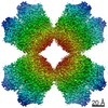
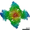
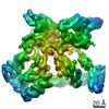
| Title | Ambient temperature structure of Bifidobacterium longum phosphoketolase with thiamine diphosphate | ||||||||||||
 Components Components | Xylulose-5-phosphate/fructose-6-phosphate phosphoketolase | ||||||||||||
 Keywords Keywords |  LYASE / XFEL-SFX / ambient temperature / aldehyde-lyase activity / carbohydrate metabolic process / thiamine-diphosphate (ThDpp) LYASE / XFEL-SFX / ambient temperature / aldehyde-lyase activity / carbohydrate metabolic process / thiamine-diphosphate (ThDpp) | ||||||||||||
| Function / homology |  Function and homology informationfructose-6-phosphate phosphoketolase / fructose-6-phosphate phosphoketolase activity / carbohydrate metabolic process / magnesium ion binding Function and homology informationfructose-6-phosphate phosphoketolase / fructose-6-phosphate phosphoketolase activity / carbohydrate metabolic process / magnesium ion bindingSimilarity search - Function | ||||||||||||
| Biological species |  Bifidobacterium longum (bacteria) Bifidobacterium longum (bacteria) | ||||||||||||
| Method | X-RAY DIFFRACTION / FREE ELECTRON LASER / MOLECULAR REPLACEMENT / Resolution: 2.5 Å | ||||||||||||
 Authors Authors | Nakata, K. / Kashiwagi, T. / Nango, E. / Miyano, H. / Mizukoshi, T. / Iwata, S. | ||||||||||||
| Funding support |  Japan, 3items Japan, 3items
| ||||||||||||
 Citation Citation | Journal: Acta Crystallogr D Struct Biol / Year: 2023 Title: Ambient temperature structure of phosphoketolase from Bifidobacterium longum determined by serial femtosecond X-ray crystallography. Authors: Nakata, K. / Kashiwagi, T. / Kunishima, N. / Naitow, H. / Matsuura, Y. / Miyano, H. / Mizukoshi, T. / Tono, K. / Yabashi, M. / Nango, E. / Iwata, S. | ||||||||||||
| History |
|
- Structure visualization
| Structure viewer | Molecule: MolmilJmol/JSmol |
|---|
- Downloads & links
-Download
| PDBx/mmCIF format | 7c8h.cif.gz | 1.2 MB | Display | PDBx/mmCIF format |
|---|---|---|---|---|
| PDB format | pdb7c8h.ent.gz | 1 MB | Display | PDB format |
| PDBx/mmJSON format | 7c8h.json.gz | Tree view | PDBx/mmJSON format | |
| Others |  Other downloads Other downloads |
-Validation report
| Arichive directory | https://data.pdbj.org/pub/pdb/validation_reports/c8/7c8hftp://data.pdbj.org/pub/pdb/validation_reports/c8/7c8h | HTTPS FTP |
|---|
-Related structure data
| Related structure data |  7c8iC  3ai7S S: Starting model for refinement C: citing same article ( |
|---|---|
| Similar structure data |

-Links
 PDBj
PDBj


- Assembly
| Deposited unit | 
| ||||||||
|---|---|---|---|---|---|---|---|---|---|
| 1 |
| ||||||||
| Unit cell |
|
-Components
-Protein , 1 types, 8 molecules ABCDEFGH
| #1: Protein | Mass: 93450.016 Da / Num. of mol.: 8 Source method: isolated from a genetically manipulated source Source: (gene. exp.) Bifidobacterium longum (bacteria) / Gene: xfp / Production host: Escherichia coli BL21(DE3) (bacteria)References: UniProt: Q6R2Q7, fructose-6-phosphate phosphoketolase |
|---|
-Non-polymers , 6 types, 1130 molecules 


| #2: Chemical | ChemComp-TPP / Thiamine pyrophosphate Mass: 425.314 Da / Num. of mol.: 8 / Source method: obtained synthetically / Formula: C12H19N4O7P2S / Feature type: SUBJECT OF INVESTIGATION Mass: 425.314 Da / Num. of mol.: 8 / Source method: obtained synthetically / Formula: C12H19N4O7P2S / Feature type: SUBJECT OF INVESTIGATION#3: Chemical | ChemComp-CA /  Mass: 40.078 Da / Num. of mol.: 8 / Source method: obtained synthetically / Formula: Ca Mass: 40.078 Da / Num. of mol.: 8 / Source method: obtained synthetically / Formula: Ca#4: Chemical | Malic acid Mass: 134.087 Da / Num. of mol.: 2 / Source method: obtained synthetically / Formula: C4H6O5 Mass: 134.087 Da / Num. of mol.: 2 / Source method: obtained synthetically / Formula: C4H6O5#5: Chemical | Malonic acid Mass: 104.061 Da / Num. of mol.: 3 / Source method: obtained synthetically / Formula: C3H4O4 Mass: 104.061 Da / Num. of mol.: 3 / Source method: obtained synthetically / Formula: C3H4O4#6: Chemical | Succinic acid Mass: 118.088 Da / Num. of mol.: 3 / Source method: obtained synthetically / Formula: C4H6O4 Mass: 118.088 Da / Num. of mol.: 3 / Source method: obtained synthetically / Formula: C4H6O4#7: Water | ChemComp-HOH / | WaterMass: 18.015 Da / Num. of mol.: 1106 / Source method: isolated from a natural source / Formula: H2O |
|---|
-Details
| Has ligand of interest | Y |
|---|---|
| Sequence details | Database reference is Q6R2Q7 which had been referred by PDB code 3AI7 according to author's request. ...Database reference is Q6R2Q7 which had been referred by PDB code 3AI7 according to author's request. Residue 575 is originally UNK. |
-Experimental details
-Experiment
| Experiment | Method: X-RAY DIFFRACTION / Number of used crystals: 1 |
|---|
- Sample preparation
| Crystal | Density Matthews: 2.89 Å3/Da / Density % sol: 57.38 % / Description: rod-like crystal |
|---|---|
| Crystal grow | Temperature: 298 K / Method: batch mode Details: Equal amount of 15 mg ml-1 PKT solution (4 mM DTT, 10 mg ml-1 ThDpp) and precipitant solution (50 mM malonate, 12% tacsimate (pH5), 17% PEG3350) were mixed in order to prepare seed crystals. ...Details: Equal amount of 15 mg ml-1 PKT solution (4 mM DTT, 10 mg ml-1 ThDpp) and precipitant solution (50 mM malonate, 12% tacsimate (pH5), 17% PEG3350) were mixed in order to prepare seed crystals. Preparations of seed crystals of PKT were performed by utilizing light induced crystallization method which promoted to form initial crystal nucleus by evanescent field arised from light irradiation to gold nano-particle vapored on general polystyrene plate (Fujifilm Wako). After 16 h, precipitated crystals were homogenized and centrifuged to use supernatant as seed solution. For the purpose of forming micro-sized crystals of PKT, 1176 micro l of protein-precipitant solution mixture as the same components of seed crystals preparation was mixed with 120 micro l of the seed solution in a 1.5 ml tube. After gentle rotation at 25 degree C for 2 h, the supernatant was exchanged to the same precipitant solution. The same buffer exchange was iteratively conducted after 10 h and 16 h. |
-Data collection
| Diffraction | Mean temperature: 293 K / Serial crystal experiment: Y |
|---|---|
| Diffraction source | Source: FREE ELECTRON LASER / Site: SACLA / Beamline: BL3 / Wavelength: 1.7714 Å |
| Detector | Type: MPCCD / Detector: CCD / Date: May 13, 2016 |
| Radiation | Protocol: SINGLE WAVELENGTH / Monochromatic (M) / Laue (L): M / Scattering type: x-ray |
| Radiation wavelength | Wavelength: 1.7714 Å / Relative weight: 1 |
| Reflection | Resolution: 2.5→46.8 Å / Num. obs: 292114 / % possible obs: 100 % / Redundancy: 234 % / Biso Wilson estimate: 57.854 Å2 / CC1/2: 0.938 / R split: 0.215 / Net I/σ(I): 3.64 |
| Reflection shell | Resolution: 2.5→2.59 Å / Redundancy: 165.2 % / Mean I/σ(I) obs: 1.11 / Num. unique obs: 29113 / CC1/2: 0.493 / R split: 1.006 / % possible all: 100 |
| Serial crystallography sample delivery | Method: injection |
- Processing
| Software |
| ||||||||||||||||||||||||||||||||||||||||||||||||||||||||||||
|---|---|---|---|---|---|---|---|---|---|---|---|---|---|---|---|---|---|---|---|---|---|---|---|---|---|---|---|---|---|---|---|---|---|---|---|---|---|---|---|---|---|---|---|---|---|---|---|---|---|---|---|---|---|---|---|---|---|---|---|---|---|
| Refinement | Method to determine structure: MOLECULAR REPLACEMENT Starting model: 3AI7 Resolution: 2.5→46.8 Å / Cor.coef. Fo:Fc: 0.97 / Cor.coef. Fo:Fc free: 0.943 / WRfactor Rfree: 0.238 / WRfactor Rwork: 0.1757 / FOM work R set: 0.7736 / SU B: 11.561 / SU ML: 0.236 / SU R Cruickshank DPI: 0.3659 / SU Rfree: 0.2414 / Cross valid method: THROUGHOUT / σ(F): 0 / ESU R: 0.366 / ESU R Free: 0.241 / Stereochemistry target values: MAXIMUM LIKELIHOOD Details: HYDROGENS HAVE BEEN ADDED IN THE RIDING POSITIONS U VALUES : REFINED INDIVIDUALLY
| ||||||||||||||||||||||||||||||||||||||||||||||||||||||||||||
| Solvent computation | Ion probe radii: 0.8 Å / Shrinkage radii: 0.8 Å / VDW probe radii: 1.2 Å / Solvent model: MASK | ||||||||||||||||||||||||||||||||||||||||||||||||||||||||||||
| Displacement parameters | Biso max: 163.71 Å2 / Biso mean: 54.255 Å2 / Biso min: 25.86 Å2
| ||||||||||||||||||||||||||||||||||||||||||||||||||||||||||||
| Refinement step | Cycle: final / Resolution: 2.5→46.8 Å
| ||||||||||||||||||||||||||||||||||||||||||||||||||||||||||||
| Refine LS restraints |
| ||||||||||||||||||||||||||||||||||||||||||||||||||||||||||||
| LS refinement shell | Resolution: 2.5→2.565 Å / Rfactor Rfree error: 0 / Total num. of bins used: 20
|
