 Movie
Movie Controller


+ Open data

- Basic information
| Entry | Database: PDB / ID: 7c61 | ||||||
|---|---|---|---|---|---|---|---|
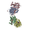
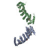
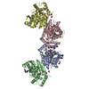
| Title | Crystal structure of 5-HT1B-BRIL and SRP2070_Fab complex | ||||||
 Components Components |
| ||||||
 Keywords Keywords |  SIGNALING PROTEIN / GPCR / BRIL / Crystallization / Antibody SIGNALING PROTEIN / GPCR / BRIL / Crystallization / Antibody | ||||||
| Function / homology |  Function and homology information Function and homology informationvoltage-gated calcium channel activity involved in regulation of presynaptic cytosolic calcium levels / adenylate cyclase-inhibiting serotonin receptor signaling pathway / negative regulation of gamma-aminobutyric acid secretion / G protein-coupled serotonin receptor complex / serotonergic synapse / regulation of behavior / Serotonin receptors / negative regulation of synaptic transmission, GABAergic / response to mineralocorticoid / negative regulation of serotonin secretion ...voltage-gated calcium channel activity involved in regulation of presynaptic cytosolic calcium levels / adenylate cyclase-inhibiting serotonin receptor signaling pathway / negative regulation of gamma-aminobutyric acid secretion / G protein-coupled serotonin receptor complex / serotonergic synapse / regulation of behavior / Serotonin receptors / negative regulation of synaptic transmission, GABAergic / response to mineralocorticoid / negative regulation of serotonin secretion / drinking behavior / cellular response to temperature stimulus / serotonin binding / bone remodeling / negative regulation of synaptic transmission, glutamatergic / G protein-coupled serotonin receptor activity / vasoconstriction / protein kinase C-activating G protein-coupled receptor signaling pathway / neurotransmitter receptor activity / G protein-coupled receptor internalization / regulation of synaptic vesicle exocytosis / regulation of dopamine secretion / heterocyclic compound binding / cellular response to alkaloid / G protein-coupled receptor signaling pathway, coupled to cyclic nucleotide second messenger / calyx of Held / positive regulation of vascular associated smooth muscle cell proliferation / adenylate cyclase-inhibiting G protein-coupled receptor signaling pathway / presynaptic modulation of chemical synaptic transmission / response to cocaine / cellular response to xenobiotic stimulus / presynaptic membrane / G alpha (i) signalling events / chemical synaptic transmission / response to ethanol / electron transfer activity / periplasmic space / iron ion binding / dendrite / heme binding / endoplasmic reticulum / plasma membraneSimilarity search - Function | ||||||
| Biological species |  Mus musculus (house mouse) Mus musculus (house mouse) Homo sapiens (human) Homo sapiens (human) Escherichia coli (E. coli) Escherichia coli (E. coli) | ||||||
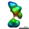
| Method | X-RAY DIFFRACTION / SYNCHROTRON / MOLECULAR REPLACEMENT / molecular replacement / Resolution: 3 Å | ||||||
 Authors Authors | Suzuki, M. / Miyagi, H. / Asada, H. / Yasunaga, M. / Suno, C. / Takahashi, Y. / Saito, J. / Iwata, S. | ||||||
 Citation Citation | Journal: Sci Rep / Year: 2020 Title: The discovery of a new antibody for BRIL-fused GPCR structure determination. Authors: Miyagi, H. / Asada, H. / Suzuki, M. / Takahashi, Y. / Yasunaga, M. / Suno, C. / Iwata, S. / Saito, J.I. | ||||||
| History |
|
- Structure visualization
| Structure viewer | Molecule: MolmilJmol/JSmol |
|---|
- Downloads & links
-Download
| PDBx/mmCIF format | 7c61.cif.gz | 313.5 KB | Display | PDBx/mmCIF format |
|---|---|---|---|---|
| PDB format | pdb7c61.ent.gz | 253.9 KB | Display | PDB format |
| PDBx/mmJSON format | 7c61.json.gz | Tree view | PDBx/mmJSON format | |
| Others |  Other downloads Other downloads |
-Validation report
| Arichive directory | https://data.pdbj.org/pub/pdb/validation_reports/c6/7c61ftp://data.pdbj.org/pub/pdb/validation_reports/c6/7c61 | HTTPS FTP |
|---|
-Related structure data
| Related structure data |  7c6aC  4iarS S: Starting model for refinement C: citing same article ( |
|---|---|
| Similar structure data |


-Links
 PDBj
PDBj


- Assembly
| Deposited unit | 
| ||||||||
|---|---|---|---|---|---|---|---|---|---|
| 1 |
| ||||||||
| Unit cell |
|
-Components
| #1: Antibody | Mass: 23681.053 Da / Num. of mol.: 1 Source method: isolated from a genetically manipulated source Source: (gene. exp.) Mus musculus (house mouse) / Production host: Mus musculus (house mouse) |
|---|---|
| #2: Antibody | Mass: 35237.762 Da / Num. of mol.: 1 Source method: isolated from a genetically manipulated source Source: (gene. exp.) Mus musculus (house mouse) / Production host: Mus musculus (house mouse) |
| #3: Protein | Mass: 45114.523 Da / Num. of mol.: 1 / Mutation: L138W,M1007W,H1102I,R1106L Source method: isolated from a genetically manipulated source Source: (gene. exp.) Homo sapiens (human), (gene. exp.) Escherichia coli (E. coli)Gene: HTR1B, HTR1DB, cybC / Production host:   Spodoptera frugiperda (fall armyworm) / References: UniProt: P28222, UniProt: P0ABE7 Spodoptera frugiperda (fall armyworm) / References: UniProt: P28222, UniProt: P0ABE7 |
| #4: Chemical | ChemComp-ERM / Ergotamine  Mass: 581.661 Da / Num. of mol.: 1 / Source method: obtained synthetically / Formula: C33H35N5O5 / Feature type: SUBJECT OF INVESTIGATION / Comment: neurotransmitter, alkaloid*YM Mass: 581.661 Da / Num. of mol.: 1 / Source method: obtained synthetically / Formula: C33H35N5O5 / Feature type: SUBJECT OF INVESTIGATION / Comment: neurotransmitter, alkaloid*YM |
| Has ligand of interest | Y |
-Experimental details
-Experiment
| Experiment | Method: X-RAY DIFFRACTION / Number of used crystals: 1 |
|---|
- Sample preparation
| Crystal | Density Matthews: 3.09 Å3/Da / Density % sol: 60.2 % |
|---|---|
| Crystal grow | Temperature: 293 K / Method: lipidic cubic phase Details: 30% PEG400, 0.4M SODIUM THIOCYANATE, 0.1M SODIUM ACETATE PH 5.5 |
-Data collection
| Diffraction | Mean temperature: 100 K / Serial crystal experiment: N | ||||||||||||||||||||||||||||||
|---|---|---|---|---|---|---|---|---|---|---|---|---|---|---|---|---|---|---|---|---|---|---|---|---|---|---|---|---|---|---|---|
| Diffraction source | Source: SYNCHROTRON / Site: SPring-8  / Beamline: BL32XU / Wavelength: 1 Å / Beamline: BL32XU / Wavelength: 1 Å | ||||||||||||||||||||||||||||||
| Detector | Type: DECTRIS PILATUS3 S 6M / Detector: PIXEL / Date: Nov 6, 2015 | ||||||||||||||||||||||||||||||
| Radiation | Protocol: SINGLE WAVELENGTH / Monochromatic (M) / Laue (L): M / Scattering type: x-ray | ||||||||||||||||||||||||||||||
| Radiation wavelength | Wavelength: 1 Å / Relative weight: 1 | ||||||||||||||||||||||||||||||
| Reflection | Resolution: 3→48.5 Å / Num. obs: 25815 / % possible obs: 100 % / Redundancy: 29.5 % / CC1/2: 0.992 / Rmerge(I) obs: 0.483 / Rpim(I) all: 0.09 / Rrim(I) all: 0.491 / Net I/σ(I): 9.2 / Num. measured all: 760396 / Scaling rejects: 271 | ||||||||||||||||||||||||||||||
| Reflection shell | Diffraction-ID: 1
|
-Phasing
| Phasing | Method: molecular replacement | |||||||||
|---|---|---|---|---|---|---|---|---|---|---|
| Phasing MR | Model details: Phaser MODE: MR_AUTO
|
- Processing
| Software |
| |||||||||||||||||||||||||||||||||||||||||||||||||||||||||||||||||||||||||||||||||||||||||||||||||||||||||||||||||||||||||||||
|---|---|---|---|---|---|---|---|---|---|---|---|---|---|---|---|---|---|---|---|---|---|---|---|---|---|---|---|---|---|---|---|---|---|---|---|---|---|---|---|---|---|---|---|---|---|---|---|---|---|---|---|---|---|---|---|---|---|---|---|---|---|---|---|---|---|---|---|---|---|---|---|---|---|---|---|---|---|---|---|---|---|---|---|---|---|---|---|---|---|---|---|---|---|---|---|---|---|---|---|---|---|---|---|---|---|---|---|---|---|---|---|---|---|---|---|---|---|---|---|---|---|---|---|---|---|---|
| Refinement | Method to determine structure: MOLECULAR REPLACEMENT Starting model: 4IAR Resolution: 3→40 Å / Cor.coef. Fo:Fc: 0.892 / Cor.coef. Fo:Fc free: 0.862 / Cross valid method: THROUGHOUT / σ(F): 0 / ESU R Free: 0.444 / Stereochemistry target values: MAXIMUM LIKELIHOOD Details: HYDROGENS HAVE BEEN ADDED IN THE RIDING POSITIONS U VALUES : WITH TLS ADDED
| |||||||||||||||||||||||||||||||||||||||||||||||||||||||||||||||||||||||||||||||||||||||||||||||||||||||||||||||||||||||||||||
| Solvent computation | Ion probe radii: 0.8 Å / Shrinkage radii: 0.8 Å / VDW probe radii: 1.2 Å / Solvent model: MASK | |||||||||||||||||||||||||||||||||||||||||||||||||||||||||||||||||||||||||||||||||||||||||||||||||||||||||||||||||||||||||||||
| Displacement parameters | Biso max: 360.78 Å2 / Biso mean: 152.781 Å2 / Biso min: 42.31 Å2
| |||||||||||||||||||||||||||||||||||||||||||||||||||||||||||||||||||||||||||||||||||||||||||||||||||||||||||||||||||||||||||||
| Refinement step | Cycle: final / Resolution: 3→40 Å
| |||||||||||||||||||||||||||||||||||||||||||||||||||||||||||||||||||||||||||||||||||||||||||||||||||||||||||||||||||||||||||||
| Refine LS restraints |
| |||||||||||||||||||||||||||||||||||||||||||||||||||||||||||||||||||||||||||||||||||||||||||||||||||||||||||||||||||||||||||||
| LS refinement shell | Resolution: 3→3.078 Å / Rfactor Rfree error: 0 / Total num. of bins used: 20
| |||||||||||||||||||||||||||||||||||||||||||||||||||||||||||||||||||||||||||||||||||||||||||||||||||||||||||||||||||||||||||||
| Refinement TLS params. | Method: refined / Refine-ID: X-RAY DIFFRACTION
| |||||||||||||||||||||||||||||||||||||||||||||||||||||||||||||||||||||||||||||||||||||||||||||||||||||||||||||||||||||||||||||
| Refinement TLS group |
|
