 Movie
Movie Controller


+ Open data

- Basic information
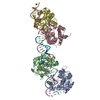
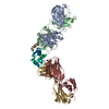
| Entry | Database: PDB / ID: 7c6a | ||||||
|---|---|---|---|---|---|---|---|
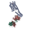
| Title | Crystal structure of AT2R-BRIL and SRP2070_Fab complex | ||||||
 Components Components |
| ||||||
 Keywords Keywords |  SIGNALING PROTEIN / GPCR / BRIL / Crystallization / Antibody SIGNALING PROTEIN / GPCR / BRIL / Crystallization / Antibody | ||||||
| Function / homology |  Function and homology information Function and homology informationangiotensin-mediated vasodilation involved in regulation of systemic arterial blood pressure / regulation of metanephros size / regulation of systemic arterial blood pressure by circulatory renin-angiotensin / angiotensin type II receptor activity / brain renin-angiotensin system / regulation of blood volume by renin-angiotensin / response to muscle activity involved in regulation of muscle adaptation / regulation of renal sodium excretion / : / type 2 angiotensin receptor binding ...angiotensin-mediated vasodilation involved in regulation of systemic arterial blood pressure / regulation of metanephros size / regulation of systemic arterial blood pressure by circulatory renin-angiotensin / angiotensin type II receptor activity / brain renin-angiotensin system / regulation of blood volume by renin-angiotensin / response to muscle activity involved in regulation of muscle adaptation / regulation of renal sodium excretion / : / type 2 angiotensin receptor binding / maintenance of blood vessel diameter homeostasis by renin-angiotensin / negative regulation of neurotrophin TRK receptor signaling pathway / positive regulation of metanephric glomerulus development / regulation of extracellular matrix assembly / receptor antagonist activity / positive regulation of activation of Janus kinase activity / regulation of renal output by angiotensin / positive regulation of NAD(P)H oxidase activity / G protein-coupled receptor signaling pathway coupled to cGMP nucleotide second messenger / renin-angiotensin regulation of aldosterone production / renal system process / positive regulation of branching involved in ureteric bud morphogenesis / positive regulation of CoA-transferase activity / positive regulation of extracellular matrix assembly / positive regulation of macrophage derived foam cell differentiation / low-density lipoprotein particle remodeling / vasoconstriction / type 1 angiotensin receptor binding / response to angiotensin / positive regulation of extrinsic apoptotic signaling pathway / exploration behavior / positive regulation of epidermal growth factor receptor signaling pathway / negative regulation of heart rate / positive regulation of cardiac muscle hypertrophy / negative regulation of blood vessel endothelial cell migration / positive regulation of gap junction assembly / positive regulation of phosphoprotein phosphatase activity / regulation of vasoconstriction / regulation of cardiac conduction / positive regulation of epithelial to mesenchymal transition / blood vessel remodeling / positive regulation of protein tyrosine kinase activity / Metabolism of Angiotensinogen to Angiotensins / nitric oxide mediated signal transduction / positive regulation of protein metabolic process / Peptide ligand-binding receptors / negative regulation of MAP kinase activity / positive regulation of endothelial cell migration / kidney development / positive regulation of nitric-oxide synthase activity / regulation of cell growth / positive regulation of cytokine production / angiotensin-activated signaling pathway / brain development / growth factor activity / serine-type endopeptidase inhibitor activity / hormone activity / negative regulation of cell growth / PPARA activates gene expression / regulation of blood pressure / positive regulation of inflammatory response / positive regulation of miRNA transcription / vasodilation / positive regulation of reactive oxygen species metabolic process / positive regulation of nitric oxide biosynthetic process / positive regulation of fibroblast proliferation / cell-cell signaling / phospholipase C-activating G protein-coupled receptor signaling pathway / positive regulation of NF-kappaB transcription factor activity / regulation of cell population proliferation / G alpha (i) signalling events / G alpha (q) signalling events / collagen-containing extracellular matrix / blood microparticle / neuron apoptotic process / regulation of apoptotic process / electron transfer activity / periplasmic space / cell surface receptor signaling pathway / positive regulation of phosphatidylinositol 3-kinase/protein kinase B signal transduction / intracellular signal transduction / inflammatory response / iron ion binding / G protein-coupled receptor signaling pathway / heme binding / positive regulation of DNA-templated transcription / extracellular space / extracellular exosome / extracellular region / plasma membrane / cytosolSimilarity search - Function | ||||||
| Biological species |  Mus musculus (house mouse) Mus musculus (house mouse) Homo sapiens (human) Homo sapiens (human) Escherichia coli (E. coli) Escherichia coli (E. coli) | ||||||
| Method | X-RAY DIFFRACTION / SYNCHROTRON / MOLECULAR REPLACEMENT / molecular replacement / Resolution: 3.4 Å | ||||||
 Authors Authors | Suzuki, M. / Miyagi, H. / Asada, H. / Yasunaga, M. / Suno, C. / Takahashi, Y. / Saito, J. / Iwata, S. | ||||||
 Citation Citation | Journal: Sci Rep / Year: 2020 Title: The discovery of a new antibody for BRIL-fused GPCR structure determination. Authors: Miyagi, H. / Asada, H. / Suzuki, M. / Takahashi, Y. / Yasunaga, M. / Suno, C. / Iwata, S. / Saito, J.I. | ||||||
| History |
|
- Structure visualization
| Structure viewer | Molecule: MolmilJmol/JSmol |
|---|
- Downloads & links
-Download
| PDBx/mmCIF format | 7c6a.cif.gz | 339.3 KB | Display | PDBx/mmCIF format |
|---|---|---|---|---|
| PDB format | pdb7c6a.ent.gz | 277.2 KB | Display | PDB format |
| PDBx/mmJSON format | 7c6a.json.gz | Tree view | PDBx/mmJSON format | |
| Others |  Other downloads Other downloads |
-Validation report
| Arichive directory | https://data.pdbj.org/pub/pdb/validation_reports/c6/7c6aftp://data.pdbj.org/pub/pdb/validation_reports/c6/7c6a | HTTPS FTP |
|---|
-Related structure data
| Related structure data |  7c61C  4zudS C: citing same article ( S: Starting model for refinement |
|---|---|
| Similar structure data |


-Links
 PDBj
PDBj


- Assembly
| Deposited unit | 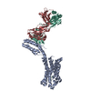
| ||||||||
|---|---|---|---|---|---|---|---|---|---|
| 1 |
| ||||||||
| Unit cell |
|
-Components
| #1: Antibody | Mass: 23681.053 Da / Num. of mol.: 1 Source method: isolated from a genetically manipulated source Source: (gene. exp.) Mus musculus (house mouse) / Production host: Mus musculus (house mouse) |
|---|---|
| #2: Antibody | Mass: 24240.152 Da / Num. of mol.: 1 Source method: isolated from a genetically manipulated source Source: (gene. exp.) Mus musculus (house mouse) / Production host: Mus musculus (house mouse) |
| #3: Protein | Mass: 48086.391 Da / Num. of mol.: 1 / Mutation: L93V, F133W, M1007W, H1102I, R1106L Source method: isolated from a genetically manipulated source Source: (gene. exp.) Homo sapiens (human), (gene. exp.) Escherichia coli (E. coli)Gene: AGTR2, cybC / Production host:   Spodoptera frugiperda (fall armyworm) / References: UniProt: P50052, UniProt: P0ABE7 Spodoptera frugiperda (fall armyworm) / References: UniProt: P50052, UniProt: P0ABE7 |
| #4: Protein/peptide | COPII Mass: 970.169 Da / Num. of mol.: 1 / Source method: obtained synthetically / Source: (synth.) Homo sapiens (human) / References: UniProt: P01019*PLUS |
| Has ligand of interest | Y |
-Experimental details
-Experiment
| Experiment | Method: X-RAY DIFFRACTION / Number of used crystals: 1 |
|---|
- Sample preparation
| Crystal | Density Matthews: 3.07 Å3/Da / Density % sol: 59.93 % |
|---|---|
| Crystal grow | Temperature: 293 K / Method: lipidic cubic phase Details: 0.05M POTASSIUM ACETATE, 0.1M MES PH6.5, 26-36% PEG300 |
-Data collection
| Diffraction | Mean temperature: 100 K / Serial crystal experiment: N | ||||||||||||||||||||||||||||||
|---|---|---|---|---|---|---|---|---|---|---|---|---|---|---|---|---|---|---|---|---|---|---|---|---|---|---|---|---|---|---|---|
| Diffraction source | Source: SYNCHROTRON / Site: SPring-8  / Beamline: BL32XU / Wavelength: 1 Å / Beamline: BL32XU / Wavelength: 1 Å | ||||||||||||||||||||||||||||||
| Detector | Type: DECTRIS PILATUS3 S 6M / Detector: PIXEL / Date: Nov 16, 2016 | ||||||||||||||||||||||||||||||
| Radiation | Protocol: SINGLE WAVELENGTH / Monochromatic (M) / Laue (L): M / Scattering type: x-ray | ||||||||||||||||||||||||||||||
| Radiation wavelength | Wavelength: 1 Å / Relative weight: 1 | ||||||||||||||||||||||||||||||
| Reflection | Resolution: 3.4→42.77 Å / Num. obs: 16178 / % possible obs: 100 % / Redundancy: 18.8 % / CC1/2: 0.985 / Rmerge(I) obs: 0.607 / Rpim(I) all: 0.14 / Rrim(I) all: 0.623 / Net I/σ(I): 5.1 / Num. measured all: 303868 / Scaling rejects: 44 | ||||||||||||||||||||||||||||||
| Reflection shell | Diffraction-ID: 1
|
-Phasing
| Phasing | Method: molecular replacement | |||||||||
|---|---|---|---|---|---|---|---|---|---|---|
| Phasing MR | Model details: Phaser MODE: MR_AUTO
|
- Processing
| Software |
| |||||||||||||||||||||||||||||||||||||||||||||||||||||||||||||||||||||||||||||||||||||||||||||||||||||||||||||||||||||||||||||
|---|---|---|---|---|---|---|---|---|---|---|---|---|---|---|---|---|---|---|---|---|---|---|---|---|---|---|---|---|---|---|---|---|---|---|---|---|---|---|---|---|---|---|---|---|---|---|---|---|---|---|---|---|---|---|---|---|---|---|---|---|---|---|---|---|---|---|---|---|---|---|---|---|---|---|---|---|---|---|---|---|---|---|---|---|---|---|---|---|---|---|---|---|---|---|---|---|---|---|---|---|---|---|---|---|---|---|---|---|---|---|---|---|---|---|---|---|---|---|---|---|---|---|---|---|---|---|
| Refinement | Method to determine structure: MOLECULAR REPLACEMENT Starting model: 4ZUD Resolution: 3.4→40 Å / Cor.coef. Fo:Fc: 0.925 / Cor.coef. Fo:Fc free: 0.869 / Cross valid method: THROUGHOUT / σ(F): 0 / ESU R Free: 0.634 / Stereochemistry target values: MAXIMUM LIKELIHOOD Details: HYDROGENS HAVE BEEN ADDED IN THE RIDING POSITIONS U VALUES : WITH TLS ADDED
| |||||||||||||||||||||||||||||||||||||||||||||||||||||||||||||||||||||||||||||||||||||||||||||||||||||||||||||||||||||||||||||
| Solvent computation | Ion probe radii: 0.8 Å / Shrinkage radii: 0.8 Å / VDW probe radii: 1.2 Å / Solvent model: MASK | |||||||||||||||||||||||||||||||||||||||||||||||||||||||||||||||||||||||||||||||||||||||||||||||||||||||||||||||||||||||||||||
| Displacement parameters | Biso max: 241.42 Å2 / Biso mean: 135.009 Å2 / Biso min: 89.75 Å2
| |||||||||||||||||||||||||||||||||||||||||||||||||||||||||||||||||||||||||||||||||||||||||||||||||||||||||||||||||||||||||||||
| Refinement step | Cycle: final / Resolution: 3.4→40 Å
| |||||||||||||||||||||||||||||||||||||||||||||||||||||||||||||||||||||||||||||||||||||||||||||||||||||||||||||||||||||||||||||
| Refine LS restraints |
| |||||||||||||||||||||||||||||||||||||||||||||||||||||||||||||||||||||||||||||||||||||||||||||||||||||||||||||||||||||||||||||
| LS refinement shell | Resolution: 3.4→3.487 Å / Rfactor Rfree error: 0 / Total num. of bins used: 20
| |||||||||||||||||||||||||||||||||||||||||||||||||||||||||||||||||||||||||||||||||||||||||||||||||||||||||||||||||||||||||||||
| Refinement TLS params. | Method: refined / Refine-ID: X-RAY DIFFRACTION
| |||||||||||||||||||||||||||||||||||||||||||||||||||||||||||||||||||||||||||||||||||||||||||||||||||||||||||||||||||||||||||||
| Refinement TLS group |
|
