 Movie
Movie Controller


+ Open data

- Basic information
| Entry | Database: PDB / ID: 7bqi | ||||||
|---|---|---|---|---|---|---|---|
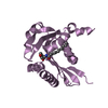
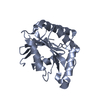
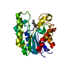
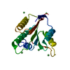
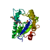
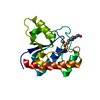
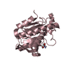
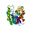
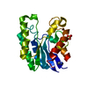
| Title | Crystal structure of FYCO1 RUN domain | ||||||
 Components Components | FYVE and coiled-coil domain-containing protein 1 | ||||||
 Keywords Keywords |  CYTOSOLIC PROTEIN / Autophagy CYTOSOLIC PROTEIN / Autophagy | ||||||
| Function / homology |  Function and homology information Function and homology informationplus-end-directed vesicle transport along microtubule / positive regulation of autophagosome maturation / autophagosome / late endosome / lysosome / intracellular membrane-bounded organelle / Golgi apparatus / membrane / metal ion bindingSimilarity search - Function | ||||||
| Biological species |  Homo sapiens (human) Homo sapiens (human) | ||||||
| Method | X-RAY DIFFRACTION / SYNCHROTRON / SIRAS / Resolution: 1.3 Å | ||||||
 Authors Authors | Sakurai, S. / Shimizu, T. / Ohto, U. | ||||||
 Citation Citation | Journal: Acta Crystallogr.,Sect.F / Year: 2020 Title: Crystal structure of the FYCO1 RUN domain suggests possible interfaces with small GTPases. Authors: Sakurai, S. / Shimizu, T. / Ohto, U. | ||||||
| History |
|
- Structure visualization
| Structure viewer | Molecule: MolmilJmol/JSmol |
|---|
- Downloads & links
-Download
| PDBx/mmCIF format | 7bqi.cif.gz | 52.7 KB | Display | PDBx/mmCIF format |
|---|---|---|---|---|
| PDB format | pdb7bqi.ent.gz | 37.1 KB | Display | PDB format |
| PDBx/mmJSON format | 7bqi.json.gz | Tree view | PDBx/mmJSON format | |
| Others |  Other downloads Other downloads |
-Validation report
| Arichive directory | https://data.pdbj.org/pub/pdb/validation_reports/bq/7bqiftp://data.pdbj.org/pub/pdb/validation_reports/bq/7bqi | HTTPS FTP |
|---|
-Related structure data
| Similar structure data |
|---|

-Links
 PDBj
PDBj

- Assembly
| Deposited unit | 
| |||||||||
|---|---|---|---|---|---|---|---|---|---|---|
| 1 |
| |||||||||
| Unit cell |
| |||||||||
| Components on special symmetry positions |
|
-Components
| #1: Protein | Mass: 20871.553 Da / Num. of mol.: 1 / Fragment: RUN domain Source method: isolated from a genetically manipulated source Source: (gene. exp.) Homo sapiens (human) / Gene: FYCO1, ZFYVE7 / Production host:  Escherichia coli (E. coli) / References: UniProt: Q9BQS8 Escherichia coli (E. coli) / References: UniProt: Q9BQS8 |
|---|---|
| #2: Water | ChemComp-HOH / Water Mass: 18.015 Da / Num. of mol.: 129 / Source method: isolated from a natural source / Formula: H2O Mass: 18.015 Da / Num. of mol.: 129 / Source method: isolated from a natural source / Formula: H2O |
-Experimental details
-Experiment
| Experiment | Method: X-RAY DIFFRACTION / Number of used crystals: 1 |
|---|
- Sample preparation
| Crystal | Density Matthews: 1.98 Å3/Da / Density % sol: 37.83 % |
|---|---|
| Crystal grow | Temperature: 277 K / Method: vapor diffusion, sitting drop / Details: 20% (w/v) PEG 3350, 180mM tri-ammonium citrate |
-Data collection
| Diffraction | Mean temperature: 100 K / Serial crystal experiment: N |
|---|---|
| Diffraction source | Source: SYNCHROTRON / Site: Photon Factory  / Beamline: AR-NE3A / Wavelength: 1 Å / Beamline: AR-NE3A / Wavelength: 1 Å |
| Detector | Type: ADSC QUANTUM 270 / Detector: CCD / Date: Nov 15, 2014 |
| Radiation | Protocol: SINGLE WAVELENGTH / Monochromatic (M) / Laue (L): M / Scattering type: x-ray |
| Radiation wavelength | Wavelength: 1 Å / Relative weight: 1 |
| Reflection | Resolution: 1.3→50 Å / Num. obs: 38506 / % possible obs: 90.7 % / Redundancy: 7.2 % / Rmerge(I) obs: 0.092 / Net I/σ(I): 41.6 |
| Reflection shell | Resolution: 1.3→1.32 Å / Rmerge(I) obs: 0.54 / Mean I/σ(I) obs: 4.5 / Num. unique obs: 1885 |
- Processing
| Software |
| ||||||||||||||||||||||||||||||||||||||||||||||||||||||||||||
|---|---|---|---|---|---|---|---|---|---|---|---|---|---|---|---|---|---|---|---|---|---|---|---|---|---|---|---|---|---|---|---|---|---|---|---|---|---|---|---|---|---|---|---|---|---|---|---|---|---|---|---|---|---|---|---|---|---|---|---|---|---|
| Refinement | Method to determine structure: SIRAS Starting model: SAD model Resolution: 1.3→45.6 Å / Cor.coef. Fo:Fc: 0.941 / Cor.coef. Fo:Fc free: 0.925 / SU B: 0.948 / SU ML: 0.041 / Cross valid method: THROUGHOUT / σ(F): 0 / ESU R: 0.067 / ESU R Free: 0.069 Details: HYDROGENS HAVE BEEN ADDED IN THE RIDING POSITIONS U VALUES : REFINED INDIVIDUALLY
| ||||||||||||||||||||||||||||||||||||||||||||||||||||||||||||
| Solvent computation | Ion probe radii: 0.8 Å / Shrinkage radii: 0.8 Å / VDW probe radii: 1.2 Å | ||||||||||||||||||||||||||||||||||||||||||||||||||||||||||||
| Displacement parameters | Biso max: 58.33 Å2 / Biso mean: 18.731 Å2 / Biso min: 8.61 Å2
| ||||||||||||||||||||||||||||||||||||||||||||||||||||||||||||
| Refinement step | Cycle: final / Resolution: 1.3→45.6 Å
| ||||||||||||||||||||||||||||||||||||||||||||||||||||||||||||
| Refine LS restraints |
| ||||||||||||||||||||||||||||||||||||||||||||||||||||||||||||
| LS refinement shell | Resolution: 1.3→1.333 Å / Rfactor Rfree error: 0 / Total num. of bins used: 20
|
