 Movie
Movie Controller

[English] 日本語
 Yorodumi
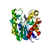
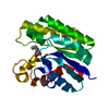
- PDB-1j00: E. coli Thioesterase I/Protease I/Lysophospholipase L1 in complex... -
+ Open data

- Basic information
| Entry | Database: PDB / ID: 1j00 | ||||||
|---|---|---|---|---|---|---|---|
| Title | E. coli Thioesterase I/Protease I/Lysophospholipase L1 in complexed with diethyl phosphono moiety | ||||||
 Components Components | Thioesterase I | ||||||
 Keywords Keywords | HYDROLASE / Protease | ||||||
| Function / homology |  Function and homology information: / : / arylesterase / phosphatidyl phospholipase B activity / lysophospholipase / : / : / palmitoyl-CoA hydrolase / fatty acyl-[ACP] hydrolase activity / : ...: / : / arylesterase / phosphatidyl phospholipase B activity / lysophospholipase / : / : / palmitoyl-CoA hydrolase / fatty acyl-[ACP] hydrolase activity / : / fatty acyl-CoA hydrolase activity / oleoyl-[acyl-carrier-protein] hydrolase / lysophospholipase activity / Hydrolases; Acting on peptide bonds (peptidases); Serine endopeptidases / arylesterase activity / lipid metabolic process / peptidase activity / outer membrane-bounded periplasmic space / periplasmic space / proteolysis / identical protein binding Function and homology information: / : / arylesterase / phosphatidyl phospholipase B activity / lysophospholipase / : / : / palmitoyl-CoA hydrolase / fatty acyl-[ACP] hydrolase activity / : ...: / : / arylesterase / phosphatidyl phospholipase B activity / lysophospholipase / : / : / palmitoyl-CoA hydrolase / fatty acyl-[ACP] hydrolase activity / : / fatty acyl-CoA hydrolase activity / oleoyl-[acyl-carrier-protein] hydrolase / lysophospholipase activity / Hydrolases; Acting on peptide bonds (peptidases); Serine endopeptidases / arylesterase activity / lipid metabolic process / peptidase activity / outer membrane-bounded periplasmic space / periplasmic space / proteolysis / identical protein bindingSimilarity search - Function | ||||||
| Biological species |  Escherichia coli (E. coli) Escherichia coli (E. coli) | ||||||
| Method | X-RAY DIFFRACTION / SYNCHROTRON / MOLECULAR REPLACEMENT / Resolution: 2 Å | ||||||
 Authors Authors | Lo, Y.-C. / Shaw, J.-F. / Liaw, Y.-C. | ||||||
 Citation Citation | Journal: J.Mol.Biol. / Year: 2003 Title: Crystal Structure of Escherichia coli Thioesterase I/Protease I/Lysophospholipase L1: Consensus Sequence Blocks Constitute the Catalytic Center of SGNH-hydrolases through a Conserved Hydrogen Bond Network Authors: Lo, Y.-C. / Lin, S.-C. / Shaw, J.-F. / Liaw, Y.-C. | ||||||
| History |
|
- Structure visualization
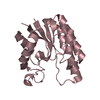
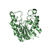
| Structure viewer | Molecule: MolmilJmol/JSmol |
|---|
- Downloads & links
-Download
| PDBx/mmCIF format | 1j00.cif.gz | 52.5 KB | Display | PDBx/mmCIF format |
|---|---|---|---|---|
| PDB format | pdb1j00.ent.gz | 36.3 KB | Display | PDB format |
| PDBx/mmJSON format | 1j00.json.gz | Tree view | PDBx/mmJSON format | |
| Others |  Other downloads Other downloads |
-Validation report
| Arichive directory | https://data.pdbj.org/pub/pdb/validation_reports/j0/1j00ftp://data.pdbj.org/pub/pdb/validation_reports/j0/1j00 | HTTPS FTP |
|---|
-Related structure data
| Related structure data |  1ivnC  1jrlSC S: Starting model for refinement C: citing same article ( |
|---|---|
| Similar structure data |


-Links
 PDBj
PDBj
- Assembly
| Deposited unit | 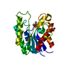
| ||||||||
|---|---|---|---|---|---|---|---|---|---|
| 1 |
| ||||||||
| 2 | 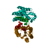
| ||||||||
| Unit cell |
|
-Components
| #1: Protein | / Acyl-CoA thioesterase I / Protease I / Lysophospholipase L1 Mass: 21697.500 Da / Num. of mol.: 1 Source method: isolated from a genetically manipulated source Source: (gene. exp.) Escherichia coli (E. coli) / Gene: tesA/apeA/pldC / Plasmid: pET-20b(+) / Production host: Escherichia coli (E. coli) / Strain (production host): BL21(DE3)References: UniProt: P29679, UniProt: P0ADA1*PLUS, Hydrolases; Acting on ester bonds; Thioester hydrolases, lysophospholipase |
|---|---|
| #2: Chemical | ChemComp-SO4 / Sulfate  Mass: 96.063 Da / Num. of mol.: 1 / Source method: obtained synthetically / Formula: SO4 Mass: 96.063 Da / Num. of mol.: 1 / Source method: obtained synthetically / Formula: SO4 |
| #3: Water | ChemComp-HOH / Water Mass: 18.015 Da / Num. of mol.: 91 / Source method: isolated from a natural source / Formula: H2O Mass: 18.015 Da / Num. of mol.: 91 / Source method: isolated from a natural source / Formula: H2O |
-Experimental details
-Experiment
| Experiment | Method: X-RAY DIFFRACTION / Number of used crystals: 1 |
|---|
- Sample preparation
| Crystal | Density Matthews: 2.47 Å3/Da / Density % sol: 50.1 % | ||||||||||||||||||||||||
|---|---|---|---|---|---|---|---|---|---|---|---|---|---|---|---|---|---|---|---|---|---|---|---|---|---|
| Crystal grow | Temperature: 298 K / Method: vapor diffusion, hanging drop / pH: 6.5 Details: 2-[N-morpholino]ethanesulfonic acid, PEGMME5000, Ammonium sulfate, pH 6.5, VAPOR DIFFUSION, HANGING DROP, temperature 298K | ||||||||||||||||||||||||
| Crystal grow | *PLUS pH: 7 / Method: unknown | ||||||||||||||||||||||||
| Components of the solutions | *PLUS
|
-Data collection
| Diffraction | Mean temperature: 133 K |
|---|---|
| Diffraction source | Source: SYNCHROTRON / Site: NSRRC  / Beamline: BL17B2 / Wavelength: 1.12714 Å / Beamline: BL17B2 / Wavelength: 1.12714 Å |
| Detector | Type: MAC Science DIP-2030 / Detector: IMAGE PLATE |
| Radiation | Monochromator: Si111 Channel / Protocol: SINGLE WAVELENGTH / Monochromatic (M) / Laue (L): M / Scattering type: x-ray |
| Radiation wavelength | Wavelength: 1.12714 Å / Relative weight: 1 |
| Reflection | Resolution: 2→27.27 Å / Num. all: 15230 / Num. obs: 14582 / % possible obs: 95.7 % / Observed criterion σ(F): 0 / Redundancy: 13.7 % / Biso Wilson estimate: 28.3 Å2 / Limit h max: 24 / Limit h min: 0 / Limit k max: 17 / Limit k min: 0 / Limit l max: 85 / Limit l min: 0 / Observed criterion F max: 1788436.9 / Observed criterion F min: 0.32 / Rmerge(I) obs: 0.04 / Net I/σ(I): 11.3 |
| Reflection shell | Resolution: 2→2.07 Å / Rmerge(I) obs: 0.381 / Mean I/σ(I) obs: 5.2 / % possible all: 98.9 |
| Reflection | *PLUS Highest resolution: 2 Å / Lowest resolution: 30 Å / Num. obs: 15230 / % possible obs: 97.7 % / Rmerge(I) obs: 0.06 |
- Processing
| Software |
| ||||||||||||||||||||||||||||||||||||||||||||||||||||||||||||||||||||||
|---|---|---|---|---|---|---|---|---|---|---|---|---|---|---|---|---|---|---|---|---|---|---|---|---|---|---|---|---|---|---|---|---|---|---|---|---|---|---|---|---|---|---|---|---|---|---|---|---|---|---|---|---|---|---|---|---|---|---|---|---|---|---|---|---|---|---|---|---|---|---|---|
| Refinement | Method to determine structure: MOLECULAR REPLACEMENT Starting model: PDB ENTRY 1JRL Resolution: 2→27.27 Å / Rfactor Rfree error: 0.008 / Occupancy max: 1 / Occupancy min: 1 / Isotropic thermal model: anisotropic / Cross valid method: THROUGHOUT / Stereochemistry target values: Engh & Huber
| ||||||||||||||||||||||||||||||||||||||||||||||||||||||||||||||||||||||
| Solvent computation | Solvent model: CNS bulk solvent model used / Bsol: 43.3515 Å2 / ksol: 0.354238 e/Å3 | ||||||||||||||||||||||||||||||||||||||||||||||||||||||||||||||||||||||
| Displacement parameters | Biso max: 120.88 Å2 / Biso mean: 51.1 Å2 / Biso min: 23.13 Å2
| ||||||||||||||||||||||||||||||||||||||||||||||||||||||||||||||||||||||
| Refine Biso |
| ||||||||||||||||||||||||||||||||||||||||||||||||||||||||||||||||||||||
| Refine analyze |
| ||||||||||||||||||||||||||||||||||||||||||||||||||||||||||||||||||||||
| Refinement step | Cycle: LAST / Resolution: 2→27.27 Å
| ||||||||||||||||||||||||||||||||||||||||||||||||||||||||||||||||||||||
| Refine LS restraints |
| ||||||||||||||||||||||||||||||||||||||||||||||||||||||||||||||||||||||
| LS refinement shell | Refine-ID: X-RAY DIFFRACTION / Total num. of bins used: 6
| ||||||||||||||||||||||||||||||||||||||||||||||||||||||||||||||||||||||
| Xplor file |
| ||||||||||||||||||||||||||||||||||||||||||||||||||||||||||||||||||||||
| Software | *PLUS Name: CNS / Classification: refinement | ||||||||||||||||||||||||||||||||||||||||||||||||||||||||||||||||||||||
| Refinement | *PLUS Highest resolution: 2 Å / Lowest resolution: 30 Å / % reflection Rfree: 10 % / Rfactor Rwork: 0.237 | ||||||||||||||||||||||||||||||||||||||||||||||||||||||||||||||||||||||
| Solvent computation | *PLUS | ||||||||||||||||||||||||||||||||||||||||||||||||||||||||||||||||||||||
| Displacement parameters | *PLUS | ||||||||||||||||||||||||||||||||||||||||||||||||||||||||||||||||||||||
| Refine LS restraints | *PLUS
| ||||||||||||||||||||||||||||||||||||||||||||||||||||||||||||||||||||||
| LS refinement shell | *PLUS Lowest resolution: 2.07 Å |
