 Movie
Movie Controller


+ Open data

- Basic information
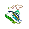
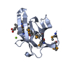
| Entry | Database: PDB / ID: 7b7j | ||||||
|---|---|---|---|---|---|---|---|
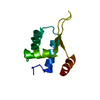
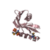
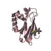
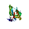
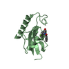
| Title | EccD5 ubiqutin like domain from Mycobacterium xenopi | ||||||
 Components Components | Secretion protein Snm4 | ||||||
 Keywords Keywords | MEMBRANE PROTEIN / ubiqutin-like cytosolic ESX secretion | ||||||
| Function / homology | Type VII secretion system membrane protein EccD / YukD-like / WXG100 protein secretion system (Wss), protein YukD / membrane => GO:0016020 / Secretion protein Snm4 Function and homology information Function and homology information | ||||||
| Biological species |  Mycobacterium xenopi (bacteria) Mycobacterium xenopi (bacteria) | ||||||
| Method | X-RAY DIFFRACTION / SYNCHROTRON / MOLECULAR REPLACEMENT / Resolution: 1.66 Å | ||||||
 Authors Authors | Beckham, K.S. / Wilmanns, M. | ||||||
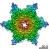
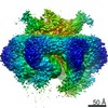
 Citation Citation | Journal: Sci Adv / Year: 2021 Title: Structure of the mycobacterial ESX-5 type VII secretion system pore complex. Authors: Katherine S H Beckham / Christina Ritter / Grzegorz Chojnowski / Daniel S Ziemianowicz / Edukondalu Mullapudi / Mandy Rettel / Mikhail M Savitski / Simon A Mortensen / Jan Kosinski / Matthias Wilmanns /  Abstract: The ESX-5 type VII secretion system is a membrane-spanning protein complex key to the virulence of mycobacterial pathogens. However, the overall architecture of the fully assembled translocation ...The ESX-5 type VII secretion system is a membrane-spanning protein complex key to the virulence of mycobacterial pathogens. However, the overall architecture of the fully assembled translocation machinery and the composition of the central secretion pore have remained unknown. Here, we present the high-resolution structure of the 2.1-megadalton ESX-5 core complex. Our structure captured a dynamic, secretion-competent conformation of the pore within a well-defined transmembrane section, sandwiched between two flexible protein layers at the cytosolic entrance and the periplasmic exit. We propose that this flexibility endows the ESX-5 machinery with large conformational plasticity required to accommodate targeted protein secretion. Compared to known secretion systems, a highly dynamic state of the pore may represent a fundamental principle of bacterial secretion machineries. | ||||||
| History |
|
- Structure visualization
| Structure viewer | Molecule: MolmilJmol/JSmol |
|---|
- Downloads & links
-Download
| PDBx/mmCIF format | 7b7j.cif.gz | 51.8 KB | Display | PDBx/mmCIF format |
|---|---|---|---|---|
| PDB format | pdb7b7j.ent.gz | 35.8 KB | Display | PDB format |
| PDBx/mmJSON format | 7b7j.json.gz | Tree view | PDBx/mmJSON format | |
| Others |  Other downloads Other downloads |
-Validation report
| Arichive directory | https://data.pdbj.org/pub/pdb/validation_reports/b7/7b7jftp://data.pdbj.org/pub/pdb/validation_reports/b7/7b7j | HTTPS FTP |
|---|
-Related structure data
| Related structure data |  7b9fC  7b9sC 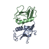 4kv2S S: Starting model for refinement C: citing same article ( |
|---|---|
| Similar structure data |


-Links
 PDBj
PDBj- Assembly
| Deposited unit | 
| ||||||||
|---|---|---|---|---|---|---|---|---|---|
| 1 |
| ||||||||
| Unit cell |
|
-Components
| #1: Protein | Mass: 14017.197 Da / Num. of mol.: 1 Source method: isolated from a genetically manipulated source Source: (gene. exp.) Mycobacterium xenopi (bacteria) / Gene: snm, NCTC10042_04572Production host: Mycolicibacterium smegmatis MC2 155 (bacteria)References: UniProt: A0A2X1TKE2 |
|---|---|
| #2: Water | ChemComp-HOH / Water Mass: 18.015 Da / Num. of mol.: 41 / Source method: isolated from a natural source / Formula: H2O Mass: 18.015 Da / Num. of mol.: 41 / Source method: isolated from a natural source / Formula: H2O |
-Experimental details
-Experiment
| Experiment | Method: X-RAY DIFFRACTION / Number of used crystals: 1 |
|---|
- Sample preparation
| Crystal grow | Temperature: 292 K / Method: vapor diffusion, sitting drop Details: 0.06 M MgCl2, CaCl2, 0.1 M Tris:bicine pH 8.5, 10 % OEG 20k, 20 % PEG MME 550 |
|---|
-Data collection
| Diffraction | Mean temperature: 100 K / Serial crystal experiment: N |
|---|---|
| Diffraction source | Source: SYNCHROTRON / Site: PETRA III, EMBL c/o DESY / Beamline: P13 (MX1) / Wavelength: 0.97 Å |
| Detector | Type: DECTRIS PILATUS3 S 6M / Detector: PIXEL / Date: Dec 21, 2017 |
| Radiation | Protocol: SINGLE WAVELENGTH / Monochromatic (M) / Laue (L): M / Scattering type: x-ray |
| Radiation wavelength | Wavelength: 0.97 Å / Relative weight: 1 |
| Reflection | Resolution: 1.66→42.57 Å / Num. obs: 10396 / % possible obs: 99.2 % / Redundancy: 6.1 % / Rmerge(I) obs: 0.049 / Net I/σ(I): 13.7 |
| Reflection shell | Resolution: 1.66→1.69 Å / Rmerge(I) obs: 1.522 / Num. unique obs: 510 / % possible all: 99.4 |
- Processing
| Software |
| ||||||||||||||||||||||||||||||||||||||||||||||||||||||||||||
|---|---|---|---|---|---|---|---|---|---|---|---|---|---|---|---|---|---|---|---|---|---|---|---|---|---|---|---|---|---|---|---|---|---|---|---|---|---|---|---|---|---|---|---|---|---|---|---|---|---|---|---|---|---|---|---|---|---|---|---|---|---|
| Refinement | Method to determine structure: MOLECULAR REPLACEMENT Starting model: 4KV2 Resolution: 1.66→30.61 Å / Cor.coef. Fo:Fc: 0.972 / Cor.coef. Fo:Fc free: 0.964 / SU B: 5.382 / SU ML: 0.087 / Cross valid method: THROUGHOUT / σ(F): 0 / ESU R: 0.101 / ESU R Free: 0.101 / Stereochemistry target values: MAXIMUM LIKELIHOOD Details: HYDROGENS HAVE BEEN ADDED IN THE RIDING POSITIONS U VALUES : WITH TLS ADDED
| ||||||||||||||||||||||||||||||||||||||||||||||||||||||||||||
| Solvent computation | Ion probe radii: 0.8 Å / Shrinkage radii: 0.8 Å / VDW probe radii: 1.2 Å / Solvent model: MASK | ||||||||||||||||||||||||||||||||||||||||||||||||||||||||||||
| Displacement parameters | Biso max: 106.07 Å2 / Biso mean: 37.551 Å2 / Biso min: 22.09 Å2
| ||||||||||||||||||||||||||||||||||||||||||||||||||||||||||||
| Refinement step | Cycle: final / Resolution: 1.66→30.61 Å
| ||||||||||||||||||||||||||||||||||||||||||||||||||||||||||||
| Refine LS restraints |
| ||||||||||||||||||||||||||||||||||||||||||||||||||||||||||||
| LS refinement shell | Resolution: 1.66→1.703 Å / Rfactor Rfree error: 0 / Total num. of bins used: 20
| ||||||||||||||||||||||||||||||||||||||||||||||||||||||||||||
| Refinement TLS params. | Method: refined / Origin x: 9.6209 Å / Origin y: 9.3155 Å / Origin z: 98.2615 Å
|
