 Movie
Movie Controller


+ Open data

- Basic information
| Entry | Database: PDB / ID: 7ah2 | ||||||||||||
|---|---|---|---|---|---|---|---|---|---|---|---|---|---|
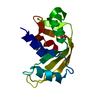
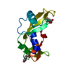
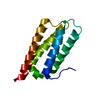
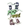
| Title | Crystal structure of Zebrafish MDM2 RING domain homodimer | ||||||||||||
 Components Components | E3 ubiquitin-protein ligase Mdm2 | ||||||||||||
 Keywords Keywords |  LIGASE / Ubiquitin ligase / RING E3 LIGASE / Ubiquitin ligase / RING E3 | ||||||||||||
| Function / homology |  Function and homology information Function and homology informationpronephric glomerulus morphogenesis / AKT phosphorylates targets in the cytosol / SUMOylation of ubiquitinylation proteins / Regulation of RUNX3 expression and activity / regulation of cell cycle => GO:0051726 / Oxidative Stress Induced Senescence / Oncogene Induced Senescence / Regulation of TP53 Activity through Phosphorylation / Regulation of TP53 Degradation / Stabilization of p53 ...pronephric glomerulus morphogenesis / AKT phosphorylates targets in the cytosol / SUMOylation of ubiquitinylation proteins / Regulation of RUNX3 expression and activity / regulation of cell cycle => GO:0051726 / Oxidative Stress Induced Senescence / Oncogene Induced Senescence / Regulation of TP53 Activity through Phosphorylation / Regulation of TP53 Degradation / Stabilization of p53 / Ub-specific processing proteases / negative regulation of intrinsic apoptotic signaling pathway by p53 class mediator / negative regulation of epithelial cell apoptotic process / response to X-ray / regulation of protein stability / RING-type E3 ubiquitin transferase / ubiquitin protein ligase activity / nucleolus / regulation of transcription by RNA polymerase II / negative regulation of apoptotic process / negative regulation of transcription by RNA polymerase II / positive regulation of transcription by RNA polymerase II / nucleoplasm / identical protein binding / metal ion binding / nucleus / cytoplasmSimilarity search - Function | ||||||||||||
| Biological species |  Danio rerio (zebrafish) Danio rerio (zebrafish) | ||||||||||||
| Method | X-RAY DIFFRACTION / SYNCHROTRON / MOLECULAR REPLACEMENT / Resolution: 2.872 Å | ||||||||||||
 Authors Authors | Magnussen, H.M. / Huang, D.T. | ||||||||||||
| Funding support |  United Kingdom, 3items United Kingdom, 3items
| ||||||||||||
 Citation Citation | Journal: J.Mol.Biol. / Year: 2021 Title: Identification of a Catalytic Active but Non-Aggregating MDM2 RING Domain Variant. Authors: Magnussen, H.M. / Huang, D.T. | ||||||||||||
| History |
|
- Structure visualization
| Structure viewer | Molecule: MolmilJmol/JSmol |
|---|
- Downloads & links
-Download
| PDBx/mmCIF format | 7ah2.cif.gz | 58.6 KB | Display | PDBx/mmCIF format |
|---|---|---|---|---|
| PDB format | pdb7ah2.ent.gz | Display | PDB format | |
| PDBx/mmJSON format | 7ah2.json.gz | Tree view | PDBx/mmJSON format | |
| Others |  Other downloads Other downloads |
-Validation report
| Arichive directory | https://data.pdbj.org/pub/pdb/validation_reports/ah/7ah2ftp://data.pdbj.org/pub/pdb/validation_reports/ah/7ah2 | HTTPS FTP |
|---|
-Related structure data
| Related structure data |  7ahyC 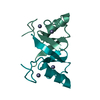 7ahzC  7ai0C 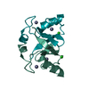 7ai1C  2vjfS S: Starting model for refinement C: citing same article ( |
|---|---|
| Similar structure data |


-Links
 PDBj
PDBj

- Assembly
| Deposited unit | 
| ||||||||
|---|---|---|---|---|---|---|---|---|---|
| 1 |
| ||||||||
| Unit cell |
|
-Components
| #1: Protein | Mass: 7798.320 Da / Num. of mol.: 2 Source method: isolated from a genetically manipulated source Source: (gene. exp.) Danio rerio (zebrafish) / Gene: mdm2 / Production host:  Escherichia coli (E. coli) Escherichia coli (E. coli)References: UniProt: O42354, RING-type E3 ubiquitin transferase #2: Chemical | ChemComp-ZN /   Mass: 65.409 Da / Num. of mol.: 4 / Source method: isolated from a natural source / Formula: Zn Mass: 65.409 Da / Num. of mol.: 4 / Source method: isolated from a natural source / Formula: Zn#3: Water | ChemComp-HOH / | Water Mass: 18.015 Da / Num. of mol.: 12 / Source method: isolated from a natural source / Formula: H2O Mass: 18.015 Da / Num. of mol.: 12 / Source method: isolated from a natural source / Formula: H2OHas ligand of interest | N | |
|---|
-Experimental details
-Experiment
| Experiment | Method: X-RAY DIFFRACTION / Number of used crystals: 1 |
|---|
- Sample preparation
| Crystal | Density Matthews: 2.25 Å3/Da / Density % sol: 45.35 % |
|---|---|
| Crystal grow | Temperature: 292 K / Method: vapor diffusion, sitting drop / pH: 6.5 Details: 0.1 M imidazole, 0.12 M monosaccharides, 37.5 % (w/v) MPD_P1K_P3350 (Morpheus, Molecular Dimensions) |
-Data collection
| Diffraction | Mean temperature: 100 K / Serial crystal experiment: N |
|---|---|
| Diffraction source | Source: SYNCHROTRON / Site: Diamond / Beamline: I04 / Wavelength: 0.9795 Å |
| Detector | Type: DECTRIS EIGER2 XE 16M / Detector: PIXEL / Date: Jun 30, 2017 |
| Radiation | Protocol: SINGLE WAVELENGTH / Monochromatic (M) / Laue (L): M / Scattering type: x-ray |
| Radiation wavelength | Wavelength: 0.9795 Å / Relative weight: 1 |
| Reflection | Resolution: 2.872→53.185 Å / Num. obs: 2814 / % possible obs: 100 % / Redundancy: 3.2 % / CC1/2: 0.949 / Net I/σ(I): 6.3 |
| Reflection shell | Resolution: 2.872→2.922 Å / Num. unique obs: 129 / CC1/2: 0.667 |
- Processing
| Software |
| |||||||||||||||||||||||||||||||||||||||||||||||||||||||||||||||||||||||||||||||||||||||||||||||||||||||||||||||||||||||||||||||||||||||||||||||||||||||||||||||||||||||||||||||||||||||||||||||||||||||||||||||||||||||||||||||||||||||
|---|---|---|---|---|---|---|---|---|---|---|---|---|---|---|---|---|---|---|---|---|---|---|---|---|---|---|---|---|---|---|---|---|---|---|---|---|---|---|---|---|---|---|---|---|---|---|---|---|---|---|---|---|---|---|---|---|---|---|---|---|---|---|---|---|---|---|---|---|---|---|---|---|---|---|---|---|---|---|---|---|---|---|---|---|---|---|---|---|---|---|---|---|---|---|---|---|---|---|---|---|---|---|---|---|---|---|---|---|---|---|---|---|---|---|---|---|---|---|---|---|---|---|---|---|---|---|---|---|---|---|---|---|---|---|---|---|---|---|---|---|---|---|---|---|---|---|---|---|---|---|---|---|---|---|---|---|---|---|---|---|---|---|---|---|---|---|---|---|---|---|---|---|---|---|---|---|---|---|---|---|---|---|---|---|---|---|---|---|---|---|---|---|---|---|---|---|---|---|---|---|---|---|---|---|---|---|---|---|---|---|---|---|---|---|---|---|---|---|---|---|---|---|---|---|---|---|---|---|---|---|---|---|
| Refinement | Method to determine structure: MOLECULAR REPLACEMENT Starting model: 2VJF Resolution: 2.872→45.21 Å / Cor.coef. Fo:Fc: 0.923 / Cor.coef. Fo:Fc free: 0.845 / SU B: 16.441 / SU ML: 0.321 / Cross valid method: FREE R-VALUE / ESU R Free: 0.477 Details: Hydrogens have been added in their riding positions
| |||||||||||||||||||||||||||||||||||||||||||||||||||||||||||||||||||||||||||||||||||||||||||||||||||||||||||||||||||||||||||||||||||||||||||||||||||||||||||||||||||||||||||||||||||||||||||||||||||||||||||||||||||||||||||||||||||||||
| Solvent computation | Ion probe radii: 0.8 Å / Shrinkage radii: 0.8 Å / VDW probe radii: 1.2 Å / Solvent model: MASK BULK SOLVENT | |||||||||||||||||||||||||||||||||||||||||||||||||||||||||||||||||||||||||||||||||||||||||||||||||||||||||||||||||||||||||||||||||||||||||||||||||||||||||||||||||||||||||||||||||||||||||||||||||||||||||||||||||||||||||||||||||||||||
| Displacement parameters | Biso mean: 23.532 Å2
| |||||||||||||||||||||||||||||||||||||||||||||||||||||||||||||||||||||||||||||||||||||||||||||||||||||||||||||||||||||||||||||||||||||||||||||||||||||||||||||||||||||||||||||||||||||||||||||||||||||||||||||||||||||||||||||||||||||||
| Refinement step | Cycle: LAST / Resolution: 2.872→45.21 Å
| |||||||||||||||||||||||||||||||||||||||||||||||||||||||||||||||||||||||||||||||||||||||||||||||||||||||||||||||||||||||||||||||||||||||||||||||||||||||||||||||||||||||||||||||||||||||||||||||||||||||||||||||||||||||||||||||||||||||
| Refine LS restraints |
| |||||||||||||||||||||||||||||||||||||||||||||||||||||||||||||||||||||||||||||||||||||||||||||||||||||||||||||||||||||||||||||||||||||||||||||||||||||||||||||||||||||||||||||||||||||||||||||||||||||||||||||||||||||||||||||||||||||||
| LS refinement shell | Refine-ID: X-RAY DIFFRACTION / Total num. of bins used: 20
|
