 Movie
Movie Controller


+ Open data

- Basic information
| Entry | Database: PDB / ID: 2y6x | ||||||
|---|---|---|---|---|---|---|---|
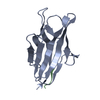
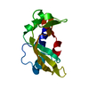
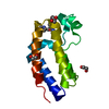
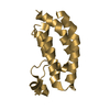
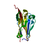
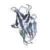
| Title | Structure of Psb27 from Thermosynechococcus elongatus | ||||||
 Components Components | PHOTOSYSTEM II 11 KD PROTEIN | ||||||
 Keywords Keywords | PHOTOSYNTHESIS | ||||||
| Function / homology |  Function and homology informationphotosystem II repair / thylakoid lumen / thylakoid / photosystem II assembly / photosystem II / plasma membrane-derived thylakoid membrane Function and homology informationphotosystem II repair / thylakoid lumen / thylakoid / photosystem II assembly / photosystem II / plasma membrane-derived thylakoid membraneSimilarity search - Function | ||||||
| Biological species |   THERMOSYNECHOCOCCUS ELONGATUS (bacteria) THERMOSYNECHOCOCCUS ELONGATUS (bacteria) | ||||||
| Method | X-RAY DIFFRACTION / SYNCHROTRON / MOLECULAR REPLACEMENT / Resolution: 1.6 Å | ||||||
 Authors Authors | Michoux, F. / Takasaka, K. / Boehm, M. / Nixon, P.J. / Murray, J.W. | ||||||
 Citation Citation | Journal: Photosynth.Res. / Year: 2012 Title: Crystal Structure of the Psb27 Assembly Factor at 1.6A: Implications for Binding to Photosystem II. Authors: Michoux, F. / Takasaka, K. / Boehm, M. / Komenda, J. / Nixon, P.J. / Murray, J.W. | ||||||
| History |
|
- Structure visualization
| Structure viewer | Molecule: MolmilJmol/JSmol |
|---|
- Downloads & links
-Download
| PDBx/mmCIF format | 2y6x.cif.gz | 62.8 KB | Display | PDBx/mmCIF format |
|---|---|---|---|---|
| PDB format | pdb2y6x.ent.gz | 46.7 KB | Display | PDB format |
| PDBx/mmJSON format | 2y6x.json.gz | Tree view | PDBx/mmJSON format | |
| Others |  Other downloads Other downloads |
-Validation report
| Arichive directory | https://data.pdbj.org/pub/pdb/validation_reports/y6/2y6xftp://data.pdbj.org/pub/pdb/validation_reports/y6/2y6x | HTTPS FTP |
|---|
-Related structure data
| Related structure data |  2kmfS S: Starting model for refinement |
|---|---|
| Similar structure data |


-Links
 PDBj
PDBj


- Assembly
| Deposited unit | 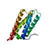
| ||||||||
|---|---|---|---|---|---|---|---|---|---|
| 1 |
| ||||||||
| Unit cell |
|
-Components
| #1: Protein | / PSB27 Mass: 12689.293 Da / Num. of mol.: 1 / Fragment: RESIDUES 24-134 Source method: isolated from a genetically manipulated source Source: (gene. exp.) THERMOSYNECHOCOCCUS ELONGATUS (bacteria)Plasmid: PRSET-A / Production host: ESCHERICHIA COLI (E. coli) / Strain (production host): K-12 / Variant (production host): KRX / References: UniProt: Q8DG60 |
|---|---|
| #2: Chemical | ChemComp-CL / Chloride  Mass: 35.453 Da / Num. of mol.: 1 / Source method: obtained synthetically / Formula: Cl Mass: 35.453 Da / Num. of mol.: 1 / Source method: obtained synthetically / Formula: Cl |
| #3: Water | ChemComp-HOH / Water Mass: 18.015 Da / Num. of mol.: 130 / Source method: isolated from a natural source / Formula: H2O Mass: 18.015 Da / Num. of mol.: 130 / Source method: isolated from a natural source / Formula: H2O |
-Experimental details
-Experiment
| Experiment | Method: X-RAY DIFFRACTION / Number of used crystals: 1 |
|---|
- Sample preparation
| Crystal | Density Matthews: 2.03 Å3/Da / Density % sol: 39.35 % / Description: NONE |
|---|---|
| Crystal grow | Method: vapor diffusion, hanging drop Details: 35% W/V PEG 4K MIXED WITH EQUAL VOLUME OF 20MG/ML PROTEIN SOLUTION, HANGING DROP VAPOUR DIFFUSION |
-Data collection
| Diffraction | Mean temperature: 100 K |
|---|---|
| Diffraction source | Source: SYNCHROTRON / Site: Diamond  / Beamline: I04 / Wavelength: 1.2831 / Beamline: I04 / Wavelength: 1.2831 |
| Detector | Type: ADSC CCD / Detector: CCD / Date: Jun 30, 2010 / Details: MIRRORS |
| Radiation | Protocol: SINGLE WAVELENGTH / Monochromatic (M) / Laue (L): M / Scattering type: x-ray |
| Radiation wavelength | Wavelength: 1.2831 Å / Relative weight: 1 |
| Reflection | Resolution: 1.6→30.31 Å / Num. obs: 13700 / % possible obs: 99.8 % / Observed criterion σ(I): 0 / Redundancy: 6.52 % / Rmerge(I) obs: 0.07 / Net I/σ(I): 8.31 |
| Reflection shell | Resolution: 1.6→1.69 Å / Redundancy: 6.61 % / Rmerge(I) obs: 0.53 / Mean I/σ(I) obs: 3.16 / % possible all: 100 |
- Processing
| Software |
| ||||||||||||||||||||||||||||||||||||||||||||||||||||||||||||||||||||||||||||||||||||||||||||||||||||||||||||||||||||||||||||||||||||||||||||||||||||||||||||||||||||||||||||||||||||||
|---|---|---|---|---|---|---|---|---|---|---|---|---|---|---|---|---|---|---|---|---|---|---|---|---|---|---|---|---|---|---|---|---|---|---|---|---|---|---|---|---|---|---|---|---|---|---|---|---|---|---|---|---|---|---|---|---|---|---|---|---|---|---|---|---|---|---|---|---|---|---|---|---|---|---|---|---|---|---|---|---|---|---|---|---|---|---|---|---|---|---|---|---|---|---|---|---|---|---|---|---|---|---|---|---|---|---|---|---|---|---|---|---|---|---|---|---|---|---|---|---|---|---|---|---|---|---|---|---|---|---|---|---|---|---|---|---|---|---|---|---|---|---|---|---|---|---|---|---|---|---|---|---|---|---|---|---|---|---|---|---|---|---|---|---|---|---|---|---|---|---|---|---|---|---|---|---|---|---|---|---|---|---|---|
| Refinement | Method to determine structure: MOLECULAR REPLACEMENT Starting model: PDB ENTRY 2KMF Resolution: 1.6→30.31 Å / Cor.coef. Fo:Fc: 0.957 / Cor.coef. Fo:Fc free: 0.931 / SU B: 4.004 / SU ML: 0.066 / Cross valid method: THROUGHOUT / ESU R: 0.105 / ESU R Free: 0.109 / Stereochemistry target values: MAXIMUM LIKELIHOOD / Details: HYDROGENS HAVE BEEN ADDED IN THE RIDING POSITIONS.
| ||||||||||||||||||||||||||||||||||||||||||||||||||||||||||||||||||||||||||||||||||||||||||||||||||||||||||||||||||||||||||||||||||||||||||||||||||||||||||||||||||||||||||||||||||||||
| Solvent computation | Ion probe radii: 0.8 Å / Shrinkage radii: 0.8 Å / VDW probe radii: 1.4 Å / Solvent model: MASK | ||||||||||||||||||||||||||||||||||||||||||||||||||||||||||||||||||||||||||||||||||||||||||||||||||||||||||||||||||||||||||||||||||||||||||||||||||||||||||||||||||||||||||||||||||||||
| Displacement parameters | Biso mean: 16.831 Å2
| ||||||||||||||||||||||||||||||||||||||||||||||||||||||||||||||||||||||||||||||||||||||||||||||||||||||||||||||||||||||||||||||||||||||||||||||||||||||||||||||||||||||||||||||||||||||
| Refinement step | Cycle: LAST / Resolution: 1.6→30.31 Å
| ||||||||||||||||||||||||||||||||||||||||||||||||||||||||||||||||||||||||||||||||||||||||||||||||||||||||||||||||||||||||||||||||||||||||||||||||||||||||||||||||||||||||||||||||||||||
| Refine LS restraints |
|