 Movie
Movie Controller

[English] 日本語
 Yorodumi
- PDB-7aag: Crystal structure of human phosphodiesterase 4D2 catalytic domain... -
+ Open data

- Basic information
| Entry | Database: PDB / ID: 7aag | ||||||
|---|---|---|---|---|---|---|---|
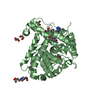
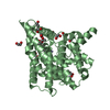
| Title | Crystal structure of human phosphodiesterase 4D2 catalytic domain with inhibitor NPD-617 | ||||||
 Components Components | cAMP-specific 3',5'-cyclic phosphodiesterase 4D | ||||||
 Keywords Keywords |  HYDROLASE / Phosphodiesterase / cAMP hydrolysis / alternative splicing HYDROLASE / Phosphodiesterase / cAMP hydrolysis / alternative splicing | ||||||
| Function / homology |  Function and homology information Function and homology informationsignaling receptor regulator activity / negative regulation of relaxation of cardiac muscle / negative regulation of heart contraction / 3',5'-cyclic-AMP phosphodiesterase / positive regulation of interleukin-5 production / regulation of cardiac muscle cell contraction / establishment of endothelial barrier / negative regulation of cAMP-mediated signaling / beta-2 adrenergic receptor binding / regulation of calcium ion transmembrane transport via high voltage-gated calcium channel ...signaling receptor regulator activity / negative regulation of relaxation of cardiac muscle / negative regulation of heart contraction / 3',5'-cyclic-AMP phosphodiesterase / positive regulation of interleukin-5 production / regulation of cardiac muscle cell contraction / establishment of endothelial barrier / negative regulation of cAMP-mediated signaling / beta-2 adrenergic receptor binding / regulation of calcium ion transmembrane transport via high voltage-gated calcium channel / positive regulation of heart rate / heterocyclic compound binding / adrenergic receptor signaling pathway / voltage-gated calcium channel complex / regulation of cell communication by electrical coupling involved in cardiac conduction / cAMP catabolic process / calcium channel regulator activity / cAMP-mediated signaling / 3',5'-cyclic-nucleotide phosphodiesterase activity / 3',5'-cyclic-AMP phosphodiesterase activity / DARPP-32 events / negative regulation of peptidyl-serine phosphorylation / regulation of release of sequestered calcium ion into cytosol by sarcoplasmic reticulum / cAMP binding / cellular response to cAMP / cellular response to epinephrine stimulus / calcium channel complex / positive regulation of interleukin-2 production / regulation of heart rate / positive regulation of type II interferon production / ATPase binding / T cell receptor signaling pathway / G alpha (s) signalling events / scaffold protein binding / transmembrane transporter binding / apical plasma membrane / centrosome / perinuclear region of cytoplasm / enzyme binding / signal transduction / membrane / metal ion binding / nucleus / plasma membrane / cytosolSimilarity search - Function | ||||||
| Biological species |  Homo sapiens (human) Homo sapiens (human) | ||||||
| Method | X-RAY DIFFRACTION / SYNCHROTRON / MOLECULAR REPLACEMENT / Resolution: 1.79 Å | ||||||
 Authors Authors | Singh, A.K. / Blaazer, A.R. / Zara, L. / de Esch, I.J.P. / Leurs, R. / Brown, D.G. | ||||||
| Funding support | European Union, 1items
| ||||||
 Citation Citation | Journal: To be published Title: hPDE4D2 structure with inhibitor NPD-617 Authors: Singh, A.K. / Brown, D.G. | ||||||
| History |
|
- Structure visualization
| Structure viewer | Molecule: MolmilJmol/JSmol |
|---|
- Downloads & links
-Download
| PDBx/mmCIF format | 7aag.cif.gz | 308.5 KB | Display | PDBx/mmCIF format |
|---|---|---|---|---|
| PDB format | pdb7aag.ent.gz | 248.3 KB | Display | PDB format |
| PDBx/mmJSON format | 7aag.json.gz | Tree view | PDBx/mmJSON format | |
| Others |  Other downloads Other downloads |
-Validation report
| Arichive directory | https://data.pdbj.org/pub/pdb/validation_reports/aa/7aagftp://data.pdbj.org/pub/pdb/validation_reports/aa/7aag | HTTPS FTP |
|---|
-Related structure data
| Related structure data |  3sl3S S: Starting model for refinement |
|---|---|
| Similar structure data |


-Links
 PDBj
PDBj


- Assembly
| Deposited unit | 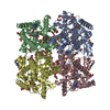
| ||||||||||||||||||||||||||||||||||||||||||||||||||||||||||||||||||||||||||||||||||||||||||||||||||||||||||||||||||||||||||||||||||||||||||||||||||||||||||||||||||||||||||||||||
|---|---|---|---|---|---|---|---|---|---|---|---|---|---|---|---|---|---|---|---|---|---|---|---|---|---|---|---|---|---|---|---|---|---|---|---|---|---|---|---|---|---|---|---|---|---|---|---|---|---|---|---|---|---|---|---|---|---|---|---|---|---|---|---|---|---|---|---|---|---|---|---|---|---|---|---|---|---|---|---|---|---|---|---|---|---|---|---|---|---|---|---|---|---|---|---|---|---|---|---|---|---|---|---|---|---|---|---|---|---|---|---|---|---|---|---|---|---|---|---|---|---|---|---|---|---|---|---|---|---|---|---|---|---|---|---|---|---|---|---|---|---|---|---|---|---|---|---|---|---|---|---|---|---|---|---|---|---|---|---|---|---|---|---|---|---|---|---|---|---|---|---|---|---|---|---|---|---|
| 1 | 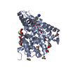
| ||||||||||||||||||||||||||||||||||||||||||||||||||||||||||||||||||||||||||||||||||||||||||||||||||||||||||||||||||||||||||||||||||||||||||||||||||||||||||||||||||||||||||||||||
| 2 | 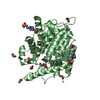
| ||||||||||||||||||||||||||||||||||||||||||||||||||||||||||||||||||||||||||||||||||||||||||||||||||||||||||||||||||||||||||||||||||||||||||||||||||||||||||||||||||||||||||||||||
| 3 | 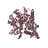
| ||||||||||||||||||||||||||||||||||||||||||||||||||||||||||||||||||||||||||||||||||||||||||||||||||||||||||||||||||||||||||||||||||||||||||||||||||||||||||||||||||||||||||||||||
| 4 | 
| ||||||||||||||||||||||||||||||||||||||||||||||||||||||||||||||||||||||||||||||||||||||||||||||||||||||||||||||||||||||||||||||||||||||||||||||||||||||||||||||||||||||||||||||||
| Unit cell |
| ||||||||||||||||||||||||||||||||||||||||||||||||||||||||||||||||||||||||||||||||||||||||||||||||||||||||||||||||||||||||||||||||||||||||||||||||||||||||||||||||||||||||||||||||
| Noncrystallographic symmetry (NCS) | NCS domain:
NCS domain segments: Component-ID: 0 / Refine code: 0
NCS ensembles :
|
-Components
-Protein , 1 types, 4 molecules ABCD
| #1: Protein | Mass: 41808.242 Da / Num. of mol.: 4 Source method: isolated from a genetically manipulated source Details: residues 381-740 / Source: (gene. exp.) Homo sapiens (human) / Gene: PDE4D, DPDE3 / Plasmid: pET15b / Production host:  Escherichia coli BL21(DE3) (bacteria) / Variant (production host): codon plus Escherichia coli BL21(DE3) (bacteria) / Variant (production host): codon plusReferences: UniProt: Q08499, 3',5'-cyclic-AMP phosphodiesterase |
|---|
-Non-polymers , 10 types, 965 molecules 


| #2: Chemical | ChemComp-ZN /  Mass: 65.409 Da / Num. of mol.: 4 / Source method: obtained synthetically / Formula: Zn Mass: 65.409 Da / Num. of mol.: 4 / Source method: obtained synthetically / Formula: Zn#3: Chemical | ChemComp-MG /  Mass: 24.305 Da / Num. of mol.: 5 / Source method: obtained synthetically / Formula: Mg Mass: 24.305 Da / Num. of mol.: 5 / Source method: obtained synthetically / Formula: Mg#4: Chemical | ChemComp-QWT /  Mass: 546.661 Da / Num. of mol.: 4 / Source method: obtained synthetically / Formula: C30H38N6O4 / Feature type: SUBJECT OF INVESTIGATION Mass: 546.661 Da / Num. of mol.: 4 / Source method: obtained synthetically / Formula: C30H38N6O4 / Feature type: SUBJECT OF INVESTIGATION#5: Chemical | ChemComp-EDO / Ethylene glycol Mass: 62.068 Da / Num. of mol.: 50 / Source method: obtained synthetically / Formula: C2H6O2 Mass: 62.068 Da / Num. of mol.: 50 / Source method: obtained synthetically / Formula: C2H6O2#6: Chemical | ChemComp-PEG / Diethylene glycol Mass: 106.120 Da / Num. of mol.: 5 / Source method: obtained synthetically / Formula: C4H10O3 Mass: 106.120 Da / Num. of mol.: 5 / Source method: obtained synthetically / Formula: C4H10O3#7: Chemical | ChemComp-DMS / Dimethyl sulfoxide Mass: 78.133 Da / Num. of mol.: 12 / Source method: obtained synthetically / Formula: C2H6OS / Comment: DMSO, precipitant*YM Mass: 78.133 Da / Num. of mol.: 12 / Source method: obtained synthetically / Formula: C2H6OS / Comment: DMSO, precipitant*YM#8: Chemical | ChemComp-EPE / HEPES Mass: 238.305 Da / Num. of mol.: 5 / Source method: obtained synthetically / Formula: C8H18N2O4S / Comment: pH buffer*YM Mass: 238.305 Da / Num. of mol.: 5 / Source method: obtained synthetically / Formula: C8H18N2O4S / Comment: pH buffer*YM#9: Chemical | ChemComp-ILE / | Isoleucine Type: L-peptide linking / Mass: 131.173 Da / Num. of mol.: 1 / Source method: obtained synthetically / Formula: C6H13NO2 Type: L-peptide linking / Mass: 131.173 Da / Num. of mol.: 1 / Source method: obtained synthetically / Formula: C6H13NO2#10: Chemical | ChemComp-PRO / | Proline Type: L-peptide linking / Mass: 115.130 Da / Num. of mol.: 1 / Source method: obtained synthetically / Formula: C5H9NO2 Type: L-peptide linking / Mass: 115.130 Da / Num. of mol.: 1 / Source method: obtained synthetically / Formula: C5H9NO2#11: Water | ChemComp-HOH / | WaterMass: 18.015 Da / Num. of mol.: 878 / Source method: isolated from a natural source / Formula: H2O |
|---|
-Details
| Has ligand of interest | Y |
|---|
-Experimental details
-Experiment
| Experiment | Method: X-RAY DIFFRACTION / Number of used crystals: 1 |
|---|
- Sample preparation
| Crystal | Density Matthews: 2.63 Å3/Da / Density % sol: 53.25 % |
|---|---|
| Crystal grow | Temperature: 293 K / Method: vapor diffusion, hanging drop / pH: 7.5 Details: 24% PEG 3350, 30% Ethylene glycol, 0.1 M HEPES pH 7.5 |
-Data collection
| Diffraction | Mean temperature: 100 K / Serial crystal experiment: N |
|---|---|
| Diffraction source | Source: SYNCHROTRON / Site: Diamond  / Beamline: I04 / Wavelength: 0.97949 Å / Beamline: I04 / Wavelength: 0.97949 Å |
| Detector | Type: DECTRIS PILATUS 6M / Detector: PIXEL / Date: Dec 7, 2015 |
| Radiation | Protocol: SINGLE WAVELENGTH / Monochromatic (M) / Laue (L): M / Scattering type: x-ray |
| Radiation wavelength | Wavelength: 0.97949 Å / Relative weight: 1 |
| Reflection | Resolution: 1.79→66.94 Å / Num. obs: 165809 / % possible obs: 100 % / Redundancy: 6.7 % / CC1/2: 0.99 / Rmerge(I) obs: 0.084 / Rpim(I) all: 0.052 / Rrim(I) all: 0.099 / Net I/σ(I): 13.5 |
| Reflection shell | Resolution: 1.79→1.84 Å / Redundancy: 6.8 % / Rmerge(I) obs: 1.26 / Mean I/σ(I) obs: 1.4 / Num. unique obs: 12112 / CC1/2: 0.52 / Rpim(I) all: 0.789 / Rrim(I) all: 1.495 / % possible all: 100 |
- Processing
| Software |
| |||||||||||||||||||||||||||||||||||||||||||||||||||||||||||||||||
|---|---|---|---|---|---|---|---|---|---|---|---|---|---|---|---|---|---|---|---|---|---|---|---|---|---|---|---|---|---|---|---|---|---|---|---|---|---|---|---|---|---|---|---|---|---|---|---|---|---|---|---|---|---|---|---|---|---|---|---|---|---|---|---|---|---|---|
| Refinement | Method to determine structure: MOLECULAR REPLACEMENT Starting model: 3SL3 Resolution: 1.79→66.94 Å / Cor.coef. Fo:Fc: 0.968 / Cor.coef. Fo:Fc free: 0.958 / SU B: 2.5 / SU ML: 0.074 / Cross valid method: THROUGHOUT / σ(F): 0 / ESU R: 0.101 / ESU R Free: 0.099 / Stereochemistry target values: MAXIMUM LIKELIHOOD Details: HYDROGENS HAVE BEEN ADDED IN THE RIDING POSITIONS U VALUES : REFINED INDIVIDUALLY
| |||||||||||||||||||||||||||||||||||||||||||||||||||||||||||||||||
| Solvent computation | Ion probe radii: 0.8 Å / Shrinkage radii: 0.8 Å / VDW probe radii: 1.2 Å / Solvent model: MASK | |||||||||||||||||||||||||||||||||||||||||||||||||||||||||||||||||
| Displacement parameters | Biso max: 115.66 Å2 / Biso mean: 30.396 Å2 / Biso min: 8.53 Å2
| |||||||||||||||||||||||||||||||||||||||||||||||||||||||||||||||||
| Refinement step | Cycle: final / Resolution: 1.79→66.94 Å
| |||||||||||||||||||||||||||||||||||||||||||||||||||||||||||||||||
| Refine LS restraints |
| |||||||||||||||||||||||||||||||||||||||||||||||||||||||||||||||||
| Refine LS restraints NCS | Refine-ID: X-RAY DIFFRACTION / Type: interatomic distance / Weight position: 0.05
| |||||||||||||||||||||||||||||||||||||||||||||||||||||||||||||||||
| LS refinement shell | Resolution: 1.79→1.836 Å / Rfactor Rfree error: 0 / Total num. of bins used: 20
|
