 Movie
Movie Controller

[English] 日本語
 Yorodumi
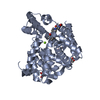
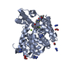
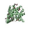
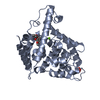
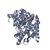
- PDB-7a2f: Crystal structure of T. brucei PDE-B1 catalytic domain with inhib... -
+ Open data

- Basic information
| Entry | Database: PDB / ID: 7a2f | ||||||
|---|---|---|---|---|---|---|---|
| Title | Crystal structure of T. brucei PDE-B1 catalytic domain with inhibitor NPD-656 | ||||||
 Components Components | Phosphodiesterase | ||||||
 Keywords Keywords | HYDROLASE / Parasitic phosphodiesterase / African trypanosomiasis / sleeping sickness | ||||||
| Function / homology |  Function and homology informationHydrolases; Acting on ester bonds; Phosphoric-diester hydrolases / axoneme / 3',5'-cyclic-nucleotide phosphodiesterase activity / 3',5'-cyclic-AMP phosphodiesterase activity / cell morphogenesis / signal transduction / metal ion binding / cytoplasm Function and homology informationHydrolases; Acting on ester bonds; Phosphoric-diester hydrolases / axoneme / 3',5'-cyclic-nucleotide phosphodiesterase activity / 3',5'-cyclic-AMP phosphodiesterase activity / cell morphogenesis / signal transduction / metal ion binding / cytoplasmSimilarity search - Function | ||||||
| Biological species |  Trypanosoma brucei (eukaryote) Trypanosoma brucei (eukaryote) | ||||||
| Method | X-RAY DIFFRACTION / SYNCHROTRON / MOLECULAR REPLACEMENT / Resolution: 2 Å | ||||||
 Authors Authors | Singh, A.K. / Blaazer, A.R. / Zara, L. / de Esch, I.J.P. / Leurs, R. / Brown, D.G. | ||||||
| Funding support | European Union, 1items
| ||||||
 Citation Citation | Journal: To be published Title: Crystal structure of T. brucei PDE-B1 catalytic domain with inhibitor NPD-656 Authors: Singh, A.K. / Brown, D.G. | ||||||
| History |
|
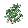
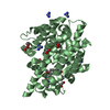
- Structure visualization
| Structure viewer | Molecule: MolmilJmol/JSmol |
|---|
- Downloads & links
-Download
| PDBx/mmCIF format | 7a2f.cif.gz | 156 KB | Display | PDBx/mmCIF format |
|---|---|---|---|---|
| PDB format | pdb7a2f.ent.gz | 119.7 KB | Display | PDB format |
| PDBx/mmJSON format | 7a2f.json.gz | Tree view | PDBx/mmJSON format | |
| Others |  Other downloads Other downloads |
-Validation report
| Arichive directory | https://data.pdbj.org/pub/pdb/validation_reports/a2/7a2fftp://data.pdbj.org/pub/pdb/validation_reports/a2/7a2f | HTTPS FTP |
|---|
-Related structure data
| Related structure data | 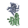 4i15S S: Starting model for refinement |
|---|---|
| Similar structure data |


-Links
 PDBj
PDBj

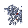
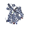
- Assembly
| Deposited unit | 
| |||||||||
|---|---|---|---|---|---|---|---|---|---|---|
| 1 | 
| |||||||||
| 2 | 
| |||||||||
| Unit cell |
| |||||||||
| Components on special symmetry positions |
|
-Components
-Protein , 1 types, 2 molecules AB
| #1: Protein | Mass: 40623.340 Da / Num. of mol.: 2 Source method: isolated from a genetically manipulated source Details: residues 565-918 / Source: (gene. exp.) Trypanosoma brucei (eukaryote) / Gene: PDEB1 / Plasmid: pET28a / Production host:  Escherichia coli BL21(DE3) (bacteria) / Variant (production host): codon plus Escherichia coli BL21(DE3) (bacteria) / Variant (production host): codon plusReferences: UniProt: Q8WQX9, Hydrolases; Acting on ester bonds; Phosphoric-diester hydrolases |
|---|
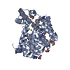
-Non-polymers , 7 types, 417 molecules 


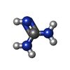

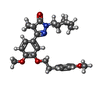

| #2: Chemical |  Mass: 65.409 Da / Num. of mol.: 2 / Source method: obtained synthetically / Formula: Zn Mass: 65.409 Da / Num. of mol.: 2 / Source method: obtained synthetically / Formula: Zn#3: Chemical |  Mass: 24.305 Da / Num. of mol.: 2 / Source method: obtained synthetically / Formula: Mg Mass: 24.305 Da / Num. of mol.: 2 / Source method: obtained synthetically / Formula: Mg#4: Chemical | Formic acid Mass: 46.025 Da / Num. of mol.: 2 / Source method: obtained synthetically / Formula: CH2O2 Mass: 46.025 Da / Num. of mol.: 2 / Source method: obtained synthetically / Formula: CH2O2#5: Chemical | ChemComp-GAI / Guanidine Mass: 59.070 Da / Num. of mol.: 7 / Source method: obtained synthetically / Formula: CH5N3 Mass: 59.070 Da / Num. of mol.: 7 / Source method: obtained synthetically / Formula: CH5N3#6: Chemical | ChemComp-GOL / Glycerol Mass: 92.094 Da / Num. of mol.: 4 / Source method: obtained synthetically / Formula: C3H8O3 Mass: 92.094 Da / Num. of mol.: 4 / Source method: obtained synthetically / Formula: C3H8O3#7: Chemical | ChemComp-QWZ / |  Mass: 464.596 Da / Num. of mol.: 1 / Source method: obtained synthetically / Formula: C28H36N2O4 / Feature type: SUBJECT OF INVESTIGATION Mass: 464.596 Da / Num. of mol.: 1 / Source method: obtained synthetically / Formula: C28H36N2O4 / Feature type: SUBJECT OF INVESTIGATION#8: Water | ChemComp-HOH / | WaterMass: 18.015 Da / Num. of mol.: 399 / Source method: isolated from a natural source / Formula: H2O |
|---|
-Details
| Has ligand of interest | Y |
|---|
-Experimental details
-Experiment
| Experiment | Method: X-RAY DIFFRACTION / Number of used crystals: 1 |
|---|
- Sample preparation
| Crystal | Density Matthews: 2.66 Å3/Da / Density % sol: 53.79 % |
|---|---|
| Crystal grow | Temperature: 277 K / Method: vapor diffusion, hanging drop / pH: 6.5 Details: 20% PEG 3350, 0.4 M sodium formate, 0.3 M guanidine, 0.1 M MES pH 6.5 |
-Data collection
| Diffraction | Mean temperature: 100 K / Serial crystal experiment: N |
|---|---|
| Diffraction source | Source: SYNCHROTRON / Site: Diamond  / Beamline: I03 / Wavelength: 0.97623 Å / Beamline: I03 / Wavelength: 0.97623 Å |
| Detector | Type: DECTRIS PILATUS3 6M / Detector: PIXEL / Date: Apr 28, 2016 / Details: CRL |
| Radiation | Protocol: SINGLE WAVELENGTH / Monochromatic (M) / Laue (L): M / Scattering type: x-ray |
| Radiation wavelength | Wavelength: 0.97623 Å / Relative weight: 1 |
| Reflection | Resolution: 2→79.38 Å / Num. obs: 54278 / % possible obs: 94.9 % / Redundancy: 2.8 % / CC1/2: 0.992 / Rmerge(I) obs: 0.091 / Rpim(I) all: 0.064 / Rrim(I) all: 0.112 / Net I/σ(I): 9.5 |
| Reflection shell | Resolution: 2→2.05 Å / Redundancy: 2.8 % / Rmerge(I) obs: 0.732 / Mean I/σ(I) obs: 2 / Num. unique obs: 4098 / CC1/2: 0.44 / Rpim(I) all: 0.532 / Rrim(I) all: 0.91 / % possible all: 97 |
- Processing
| Software |
| ||||||||||||||||||||||||||||||||||||||||||||||||||||||||||||
|---|---|---|---|---|---|---|---|---|---|---|---|---|---|---|---|---|---|---|---|---|---|---|---|---|---|---|---|---|---|---|---|---|---|---|---|---|---|---|---|---|---|---|---|---|---|---|---|---|---|---|---|---|---|---|---|---|---|---|---|---|---|
| Refinement | Method to determine structure: MOLECULAR REPLACEMENT Starting model: 4I15 Resolution: 2→79.38 Å / Cor.coef. Fo:Fc: 0.958 / Cor.coef. Fo:Fc free: 0.94 / SU B: 6.598 / SU ML: 0.159 / Cross valid method: THROUGHOUT / σ(F): 0 / ESU R: 0.174 / ESU R Free: 0.155 / Stereochemistry target values: MAXIMUM LIKELIHOOD Details: HYDROGENS HAVE BEEN ADDED IN THE RIDING POSITIONS U VALUES : REFINED INDIVIDUALLY
| ||||||||||||||||||||||||||||||||||||||||||||||||||||||||||||
| Solvent computation | Ion probe radii: 0.8 Å / Shrinkage radii: 0.8 Å / VDW probe radii: 1.2 Å / Solvent model: MASK | ||||||||||||||||||||||||||||||||||||||||||||||||||||||||||||
| Displacement parameters | Biso max: 125.3 Å2 / Biso mean: 29.834 Å2 / Biso min: 6.31 Å2
| ||||||||||||||||||||||||||||||||||||||||||||||||||||||||||||
| Refinement step | Cycle: final / Resolution: 2→79.38 Å
| ||||||||||||||||||||||||||||||||||||||||||||||||||||||||||||
| Refine LS restraints |
| ||||||||||||||||||||||||||||||||||||||||||||||||||||||||||||
| LS refinement shell | Resolution: 2→2.052 Å / Rfactor Rfree error: 0 / Total num. of bins used: 20
|
