 Movie
Movie Controller

[English] 日本語
 Yorodumi
- PDB-6ysw: E. coli anaerobic trifunctional enzyme subunit-alpha in complex w... -
+ Open data

- Basic information
| Entry | Database: PDB / ID: 6ysw | ||||||
|---|---|---|---|---|---|---|---|
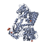
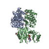
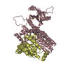
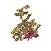
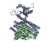
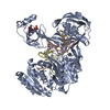
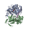
| Title | E. coli anaerobic trifunctional enzyme subunit-alpha in complex with coenzyme A | ||||||
 Components Components | Fatty acid oxidation complex subunit alpha Beta oxidation Beta oxidation | ||||||
 Keywords Keywords | OXIDOREDUCTASE / fatty acid oxidation / lipid metabolism / hydratase / dehydrogenase / trifunctional enzyme / beta oxidation | ||||||
| Function / homology |  Function and homology information Function and homology informationfatty acid beta-oxidation multienzyme complex / 3-hydroxybutyryl-CoA epimerase / 3-hydroxybutyryl-CoA epimerase activity / long-chain-3-hydroxyacyl-CoA dehydrogenase activity / 3-hydroxyacyl-CoA dehydrogenase / enoyl-CoA hydratase / 3-hydroxyacyl-CoA dehydrogenase activity / enoyl-CoA hydratase activity / fatty acid beta-oxidation / NAD+ binding / cytoplasmSimilarity search - Function | ||||||
| Biological species |  Escherichia coli (E. coli) Escherichia coli (E. coli) | ||||||
| Method | X-RAY DIFFRACTION / SYNCHROTRON / MOLECULAR REPLACEMENT / Resolution: 2.82 Å | ||||||
 Authors Authors | Sah-Teli, S.K. / Hynonen, M.J. / Wierenga, R.K. / Venkatesan, R. | ||||||
| Funding support |  Finland, 1items Finland, 1items
| ||||||
 Citation Citation | Journal: Structure / Year: 2023 Title: Structural basis for different membrane-binding properties of E. coli anaerobic and human mitochondrial beta-oxidation trifunctional enzymes Authors: Sah-Teli, S.K. / Pinkas, M. / Hynonen, M.J. / Butcher, S.J. / Wierenga, R.K. / Novacek, J. / Venkatesan, R. #1: Journal: Biochem J / Year: 2019Title: Complementary substrate specificity and distinct quaternary assembly of the aerobic and anaerobic β-oxidation trifunctional enzyme complexes. Authors: Shiv K Sah-Teli / Mikko J Hynönen / Werner Schmitz / James A Geraets / Jani Seitsonen / Jan Skov Pedersen / Sarah J Butcher / Rik K Wierenga / Rajaram Venkatesan /   Abstract: The trifunctional enzyme (TFE) catalyzes the last three steps of the fatty acid β-oxidation cycle. Two TFEs are present in , EcTFE and anEcTFE. EcTFE is expressed only under aerobic conditions, ...The trifunctional enzyme (TFE) catalyzes the last three steps of the fatty acid β-oxidation cycle. Two TFEs are present in , EcTFE and anEcTFE. EcTFE is expressed only under aerobic conditions, whereas anEcTFE is expressed also under anaerobic conditions, with nitrate or fumarate as the ultimate electron acceptor. The anEcTFE subunits have higher sequence identity with the human mitochondrial TFE (HsTFE) than with the soluble EcTFE. Like HsTFE, here it is found that anEcTFE is a membrane-bound complex. Systematic enzyme kinetic studies show that anEcTFE has a preference for medium- and long-chain enoyl-CoAs, similar to HsTFE, whereas EcTFE prefers short chain enoyl-CoA substrates. The biophysical characterization of anEcTFE and EcTFE shows that EcTFE is heterotetrameric, whereas anEcTFE is purified as a complex of two heterotetrameric units, like HsTFE. The tetrameric assembly of anEcTFE resembles the HsTFE tetramer, although the arrangement of the two anEcTFE tetramers in the octamer is different from the HsTFE octamer. These studies demonstrate that EcTFE and anEcTFE have complementary substrate specificities, allowing for complete degradation of long-chain enoyl-CoAs under aerobic conditions. The new data agree with the notion that anEcTFE and HsTFE are evolutionary closely related, whereas EcTFE belongs to a separate subfamily. | ||||||
| History |
|
- Structure visualization
| Structure viewer | Molecule: MolmilJmol/JSmol |
|---|
- Downloads & links
-Download
| PDBx/mmCIF format | 6ysw.cif.gz | 642 KB | Display | PDBx/mmCIF format |
|---|---|---|---|---|
| PDB format | pdb6ysw.ent.gz | 442.7 KB | Display | PDB format |
| PDBx/mmJSON format | 6ysw.json.gz | Tree view | PDBx/mmJSON format | |
| Others |  Other downloads Other downloads |
-Validation report
| Arichive directory | https://data.pdbj.org/pub/pdb/validation_reports/ys/6yswftp://data.pdbj.org/pub/pdb/validation_reports/ys/6ysw | HTTPS FTP |
|---|
-Related structure data
| Related structure data |  6ysvSC S: Starting model for refinement C: citing same article ( |
|---|---|
| Similar structure data |


-Links
 PDBj
PDBj


- Assembly
| Deposited unit | 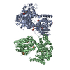
| ||||||||||||
|---|---|---|---|---|---|---|---|---|---|---|---|---|---|
| 1 | 
| ||||||||||||
| 2 | 
| ||||||||||||
| Unit cell |
| ||||||||||||
| Components on special symmetry positions |
|
-Components
| #1: Protein | Beta oxidation Mass: 78787.836 Da / Num. of mol.: 2 Source method: isolated from a genetically manipulated source Details: N-terminal 6XHis-tag Source: (gene. exp.) Escherichia coli (strain K12) (bacteria)Gene: fadJ, yfcX, b2341, JW2338 / Variant: MG1655 / Plasmid: pETDuet-1 / Cell line (production host): BL21DE3 pLys / Production host: Escherichia coli (E. coli)References: UniProt: P77399, enoyl-CoA hydratase, 3-hydroxybutyryl-CoA epimerase, 3-hydroxyacyl-CoA dehydrogenase#2: Chemical | ChemComp-SO4 / Sulfate  Mass: 96.063 Da / Num. of mol.: 13 / Source method: obtained synthetically / Formula: SO4 Mass: 96.063 Da / Num. of mol.: 13 / Source method: obtained synthetically / Formula: SO4#3: Chemical | ChemComp-COA / | Coenzyme A  Mass: 767.534 Da / Num. of mol.: 1 / Source method: obtained synthetically / Formula: C21H36N7O16P3S / Feature type: SUBJECT OF INVESTIGATION Mass: 767.534 Da / Num. of mol.: 1 / Source method: obtained synthetically / Formula: C21H36N7O16P3S / Feature type: SUBJECT OF INVESTIGATION#4: Water | ChemComp-HOH / | Water Mass: 18.015 Da / Num. of mol.: 19 / Source method: isolated from a natural source / Formula: H2O Mass: 18.015 Da / Num. of mol.: 19 / Source method: isolated from a natural source / Formula: H2OHas ligand of interest | Y | |
|---|
-Experimental details
-Experiment
| Experiment | Method: X-RAY DIFFRACTION / Number of used crystals: 1 |
|---|
- Sample preparation
| Crystal | Density Matthews: 3.1 Å3/Da / Density % sol: 60 % |
|---|---|
| Crystal grow | Temperature: 277 K / Method: vapor diffusion, sitting drop / pH: 8 Details: 100 mM Tris, 1.8 M ammonium sulfate, 4.5 % glycerol, pH 8.0, and 5 % 1,4-dioxane Temp details: cold room |
-Data collection
| Diffraction | Mean temperature: 100 K / Ambient temp details: liquid nitrogen / Serial crystal experiment: N |
|---|---|
| Diffraction source | Source: SYNCHROTRON / Site: Diamond  / Beamline: I24 / Wavelength: 0.9688 Å / Beamline: I24 / Wavelength: 0.9688 Å |
| Detector | Type: DECTRIS PILATUS 6M / Detector: PIXEL / Date: May 4, 2019 |
| Radiation | Protocol: SINGLE WAVELENGTH / Monochromatic (M) / Laue (L): M / Scattering type: x-ray |
| Radiation wavelength | Wavelength: 0.9688 Å / Relative weight: 1 |
| Reflection | Resolution: 2.82→107.67 Å / Num. obs: 28799 / % possible obs: 84.5 % / Redundancy: 4.1 % / Biso Wilson estimate: 62.5 Å2 / CC1/2: 1 / Rpim(I) all: 0.094 / Net I/σ(I): 4.8 |
| Reflection shell | Resolution: 2.82→3.15 Å / Redundancy: 4.6 % / Mean I/σ(I) obs: 1.6 / Num. unique obs: 1440 / CC1/2: 0.6 / Rpim(I) all: 0.515 / % possible all: 61.5 |
- Processing
| Software |
| |||||||||||||||||||||||||||||||||||||||||||||||||||||||||||||||||||||||||||||||||||||||||||||||||||||||||||||||||||||||||||||||||||||||||||||||||||||||||||||||||||||||||||||||
|---|---|---|---|---|---|---|---|---|---|---|---|---|---|---|---|---|---|---|---|---|---|---|---|---|---|---|---|---|---|---|---|---|---|---|---|---|---|---|---|---|---|---|---|---|---|---|---|---|---|---|---|---|---|---|---|---|---|---|---|---|---|---|---|---|---|---|---|---|---|---|---|---|---|---|---|---|---|---|---|---|---|---|---|---|---|---|---|---|---|---|---|---|---|---|---|---|---|---|---|---|---|---|---|---|---|---|---|---|---|---|---|---|---|---|---|---|---|---|---|---|---|---|---|---|---|---|---|---|---|---|---|---|---|---|---|---|---|---|---|---|---|---|---|---|---|---|---|---|---|---|---|---|---|---|---|---|---|---|---|---|---|---|---|---|---|---|---|---|---|---|---|---|---|---|---|---|
| Refinement | Method to determine structure: MOLECULAR REPLACEMENT Starting model: 6YSV Resolution: 2.82→85.72 Å / SU ML: 0.3369 / Cross valid method: FREE R-VALUE / σ(F): 1.35 / Phase error: 29.5865
| |||||||||||||||||||||||||||||||||||||||||||||||||||||||||||||||||||||||||||||||||||||||||||||||||||||||||||||||||||||||||||||||||||||||||||||||||||||||||||||||||||||||||||||||
| Solvent computation | Shrinkage radii: 0.9 Å / VDW probe radii: 1.11 Å | |||||||||||||||||||||||||||||||||||||||||||||||||||||||||||||||||||||||||||||||||||||||||||||||||||||||||||||||||||||||||||||||||||||||||||||||||||||||||||||||||||||||||||||||
| Displacement parameters | Biso mean: 58.2 Å2 | |||||||||||||||||||||||||||||||||||||||||||||||||||||||||||||||||||||||||||||||||||||||||||||||||||||||||||||||||||||||||||||||||||||||||||||||||||||||||||||||||||||||||||||||
| Refinement step | Cycle: LAST / Resolution: 2.82→85.72 Å
| |||||||||||||||||||||||||||||||||||||||||||||||||||||||||||||||||||||||||||||||||||||||||||||||||||||||||||||||||||||||||||||||||||||||||||||||||||||||||||||||||||||||||||||||
| Refine LS restraints |
| |||||||||||||||||||||||||||||||||||||||||||||||||||||||||||||||||||||||||||||||||||||||||||||||||||||||||||||||||||||||||||||||||||||||||||||||||||||||||||||||||||||||||||||||
| LS refinement shell |
| |||||||||||||||||||||||||||||||||||||||||||||||||||||||||||||||||||||||||||||||||||||||||||||||||||||||||||||||||||||||||||||||||||||||||||||||||||||||||||||||||||||||||||||||
| Refinement TLS params. | Method: refined / Refine-ID: X-RAY DIFFRACTION
| |||||||||||||||||||||||||||||||||||||||||||||||||||||||||||||||||||||||||||||||||||||||||||||||||||||||||||||||||||||||||||||||||||||||||||||||||||||||||||||||||||||||||||||||
| Refinement TLS group |
|
