 Movie
Movie Controller


+ Open data

- Basic information
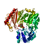
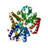
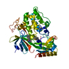
| Entry | Database: PDB / ID: 4g0b | ||||||
|---|---|---|---|---|---|---|---|
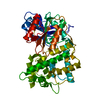
| Title | Structure of native HCT from Coffea canephora | ||||||
 Components Components | Hydroxycinnamoyl-CoA shikimate/quinate hydroxycinnamoyltransferase | ||||||
 Keywords Keywords |  TRANSFERASE / BAHD Superfamily / hydroxycinnamoyl transferase TRANSFERASE / BAHD Superfamily / hydroxycinnamoyl transferase | ||||||
| Function / homology | Transferase family / Chloramphenicol Acetyltransferase / Chloramphenicol acetyltransferase-like domain / acyltransferase activity, transferring groups other than amino-acyl groups / Chloramphenicol acetyltransferase-like domain superfamily / 2-Layer Sandwich / Alpha Beta / Hydroxycinnamoyl-CoA shikimate/quinate hydroxycinnamoyltransferase Function and homology information Function and homology information | ||||||
| Biological species |  Coffea canephora (robusta coffee) Coffea canephora (robusta coffee) | ||||||
| Method | X-RAY DIFFRACTION / SYNCHROTRON / MOLECULAR REPLACEMENT / Resolution: 3 Å | ||||||
 Authors Authors | Lallemand, L.A. / McCarthy, J.G. / McCarthy, A.A. | ||||||
 Citation Citation | Journal: Plant Physiol. / Year: 2012 Title: A structural basis for the biosynthesis of the major chlorogenic acids found in coffee. Authors: Lallemand, L.A. / Zubieta, C. / Lee, S.G. / Wang, Y. / Acajjaoui, S. / Timmins, J. / McSweeney, S. / Jez, J.M. / McCarthy, J.G. / McCarthy, A.A. | ||||||
| History |
|
- Structure visualization
| Structure viewer | Molecule: MolmilJmol/JSmol |
|---|
- Downloads & links
-Download
| PDBx/mmCIF format | 4g0b.cif.gz | 328.5 KB | Display | PDBx/mmCIF format |
|---|---|---|---|---|
| PDB format | pdb4g0b.ent.gz | 270.5 KB | Display | PDB format |
| PDBx/mmJSON format | 4g0b.json.gz | Tree view | PDBx/mmJSON format | |
| Others |  Other downloads Other downloads |
-Validation report
| Arichive directory | https://data.pdbj.org/pub/pdb/validation_reports/g0/4g0bftp://data.pdbj.org/pub/pdb/validation_reports/g0/4g0b | HTTPS FTP |
|---|
-Related structure data
| Related structure data | 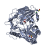 4g22C 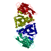 4g2mC 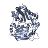 2bghS C: citing same article ( S: Starting model for refinement |
|---|---|
| Similar structure data |


-Links
 PDBj
PDBj

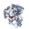
- Assembly
| Deposited unit | 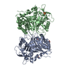
| ||||||||||||||||||
|---|---|---|---|---|---|---|---|---|---|---|---|---|---|---|---|---|---|---|---|
| 1 | 
| ||||||||||||||||||
| 2 | 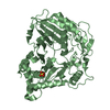
| ||||||||||||||||||
| 3 | 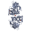
| ||||||||||||||||||
| 4 | 
| ||||||||||||||||||
| Unit cell |
| ||||||||||||||||||
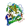
| Noncrystallographic symmetry (NCS) | NCS domain:
NCS domain segments: Component-ID: 0 / Ens-ID: 1 / Beg auth comp-ID: MET / Beg label comp-ID: MET / End auth comp-ID: ILE / End label comp-ID: ILE / Refine code: 0 / Auth seq-ID: 1 - 434 / Label seq-ID: 3 - 436
|
-Components
| #1: Protein | Mass: 48241.852 Da / Num. of mol.: 2 Source method: isolated from a genetically manipulated source Source: (gene. exp.) Coffea canephora (robusta coffee) / Gene: HCT / Plasmid: pProEX_HTb / Production host:  Escherichia coli (E. coli) / Strain (production host): BL21 Star (DE3) pLysS Escherichia coli (E. coli) / Strain (production host): BL21 Star (DE3) pLysSReferences: UniProt: A4ZKE4, shikimate O-hydroxycinnamoyltransferase#2: Chemical | ChemComp-SO4 / Sulfate  Mass: 96.063 Da / Num. of mol.: 5 / Source method: obtained synthetically / Formula: SO4 Mass: 96.063 Da / Num. of mol.: 5 / Source method: obtained synthetically / Formula: SO4#3: Water | ChemComp-HOH / | Water Mass: 18.015 Da / Num. of mol.: 28 / Source method: isolated from a natural source / Formula: H2O Mass: 18.015 Da / Num. of mol.: 28 / Source method: isolated from a natural source / Formula: H2O |
|---|
-Experimental details
-Experiment
| Experiment | Method: X-RAY DIFFRACTION / Number of used crystals: 1 |
|---|
- Sample preparation
| Crystal | Density Matthews: 2.78 Å3/Da / Density % sol: 55.68 % |
|---|---|
| Crystal grow | Temperature: 293 K / Method: vapor diffusion, sitting drop / pH: 6.5 Details: 1.6 M magnesium sulfate and 0.1 M MES, VAPOR DIFFUSION, SITTING DROP, temperature 293K, pH 6.5 |
-Data collection
| Diffraction | Mean temperature: 100 K |
|---|---|
| Diffraction source | Source: SYNCHROTRON / Site: ESRF  / Beamline: ID23-2 / Wavelength: 0.873 Å / Beamline: ID23-2 / Wavelength: 0.873 Å |
| Detector | Type: RAYONIX MX-225 / Detector: CCD / Date: May 5, 2008 / Details: KB-mirror system |
| Radiation | Monochromator: Si111 / Protocol: SINGLE WAVELENGTH / Monochromatic (M) / Laue (L): M / Scattering type: x-ray |
| Radiation wavelength | Wavelength: 0.873 Å / Relative weight: 1 |
| Reflection | Resolution: 3→50 Å / Num. all: 72116 / Num. obs: 22073 / % possible obs: 99 % / Observed criterion σ(F): 1 / Observed criterion σ(I): 1 / Rmerge(I) obs: 0.166 / Net I/σ(I): 8.5 |
| Reflection shell | Resolution: 3→3.1 Å / Rmerge(I) obs: 0.566 / Mean I/σ(I) obs: 3.1 / Num. unique all: 10301 / % possible all: 98.4 |
- Processing
| Software |
| |||||||||||||||||||||||||||||||||||||||||||||||||||||||||||||||||||||||||||
|---|---|---|---|---|---|---|---|---|---|---|---|---|---|---|---|---|---|---|---|---|---|---|---|---|---|---|---|---|---|---|---|---|---|---|---|---|---|---|---|---|---|---|---|---|---|---|---|---|---|---|---|---|---|---|---|---|---|---|---|---|---|---|---|---|---|---|---|---|---|---|---|---|---|---|---|---|
| Refinement | Method to determine structure: MOLECULAR REPLACEMENT Starting model: PDB ENTRY 2BGH Resolution: 3→50 Å / Cor.coef. Fo:Fc: 0.924 / Cor.coef. Fo:Fc free: 0.879 / SU B: 38.688 / SU ML: 0.323 / Cross valid method: THROUGHOUT / σ(F): 1 / ESU R Free: 0.43 / Stereochemistry target values: MAXIMUM LIKELIHOOD
| |||||||||||||||||||||||||||||||||||||||||||||||||||||||||||||||||||||||||||
| Solvent computation | Ion probe radii: 0.8 Å / Shrinkage radii: 0.8 Å / VDW probe radii: 1.2 Å / Solvent model: MASK | |||||||||||||||||||||||||||||||||||||||||||||||||||||||||||||||||||||||||||
| Displacement parameters | Biso mean: 27.385 Å2
| |||||||||||||||||||||||||||||||||||||||||||||||||||||||||||||||||||||||||||
| Refinement step | Cycle: LAST / Resolution: 3→50 Å
| |||||||||||||||||||||||||||||||||||||||||||||||||||||||||||||||||||||||||||
| Refine LS restraints |
| |||||||||||||||||||||||||||||||||||||||||||||||||||||||||||||||||||||||||||
| Refine LS restraints NCS | Ens-ID: 1 / Number: 465 / Refine-ID: X-RAY DIFFRACTION / Type: interatomic distance / Rms dev position: 0.22 Å / Weight position: 0.05
| |||||||||||||||||||||||||||||||||||||||||||||||||||||||||||||||||||||||||||
| LS refinement shell | Resolution: 3→3.078 Å / Total num. of bins used: 20
| |||||||||||||||||||||||||||||||||||||||||||||||||||||||||||||||||||||||||||
| Refinement TLS params. | Method: refined / Refine-ID: X-RAY DIFFRACTION
| |||||||||||||||||||||||||||||||||||||||||||||||||||||||||||||||||||||||||||
| Refinement TLS group |
|
