 Movie
Movie Controller

[English] 日本語
 Yorodumi
- PDB-6wpm: Crystal structure of a putative oligosaccharide periplasmic-bindi... -
+ Open data

- Basic information
| Entry | Database: PDB / ID: 6wpm | |||||||||
|---|---|---|---|---|---|---|---|---|---|---|
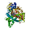
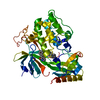
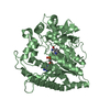
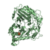
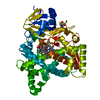
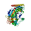
| Title | Crystal structure of a putative oligosaccharide periplasmic-binding protein from Synechococcus sp. MITs9220 in complex with zinc | |||||||||
 Components Components | Substrate-binding protein | |||||||||
 Keywords Keywords |  TRANSPORT PROTEIN / Substrate-binding protein / marine cyanobacteria / sugar-binding protein TRANSPORT PROTEIN / Substrate-binding protein / marine cyanobacteria / sugar-binding protein | |||||||||
| Biological species |  Synechococcus sp. (bacteria) Synechococcus sp. (bacteria) | |||||||||
| Method | X-RAY DIFFRACTION / SYNCHROTRON / SAD / Resolution: 2.88 Å | |||||||||
 Authors Authors | Ford, B.A. / Michie, K.A. / Paulsen, I.T. / Mabbutt, B.C. / Shah, B.S. | |||||||||
| Funding support |  Australia, 2items Australia, 2items
| |||||||||
 Citation Citation | Journal: Sci Rep / Year: 2022 Title: Novel functional insights into a modified sugar-binding protein from Synechococcus MITS9220. Authors: Ford, B.A. / Michie, K.A. / Paulsen, I.T. / Mabbutt, B.C. / Shah, B.S. #1: Journal: Acta Crystallogr. D Biol. Crystallogr. / Year: 2012 Title: Towards automated crystallographic structure refinement with phenix.refine. Authors: Afonine, P.V. / Grosse-Kunstleve, R.W. / Echols, N. / Headd, J.J. / Moriarty, N.W. / Mustyakimov, M. / Terwilliger, T.C. / Urzhumtsev, A. / Zwart, P.H. / Adams, P.D. | |||||||||
| History |
|
- Structure visualization
| Structure viewer | Molecule:  MolmilJmol/JSmol MolmilJmol/JSmol |
|---|
- Downloads & links
-Download
| PDBx/mmCIF format | 6wpm.cif.gz | 279.3 KB | Display | PDBx/mmCIF format |
|---|---|---|---|---|
| PDB format | pdb6wpm.ent.gz | 203.4 KB | Display | PDB format |
| PDBx/mmJSON format | 6wpm.json.gz | Tree view | PDBx/mmJSON format | |
| Others |  Other downloads Other downloads |
-Validation report
| Arichive directory | https://data.pdbj.org/pub/pdb/validation_reports/wp/6wpmftp://data.pdbj.org/pub/pdb/validation_reports/wp/6wpm | HTTPS FTP |
|---|
-Related structure data


-Links
 PDBj
PDBj- Assembly
| Deposited unit | 
| ||||||||||||
|---|---|---|---|---|---|---|---|---|---|---|---|---|---|
| 1 |
| ||||||||||||
| Unit cell |
| ||||||||||||
| Components on special symmetry positions |
|
-Components
-Protein , 1 types, 1 molecules A
| #1: Protein | Mass: 47341.387 Da / Num. of mol.: 1 Source method: isolated from a genetically manipulated source Source: (gene. exp.) Synechococcus sp. (bacteria) / Strain: MITs9220 / Gene: 00121 / Plasmid: pET15b / Production host: Escherichia coli BL21 (bacteria) |
|---|
-Non-polymers , 5 types, 20 molecules 


| #2: Chemical | ChemComp-GOL / Glycerol Mass: 92.094 Da / Num. of mol.: 1 / Source method: obtained synthetically / Formula: C3H8O3 Mass: 92.094 Da / Num. of mol.: 1 / Source method: obtained synthetically / Formula: C3H8O3 | ||||
|---|---|---|---|---|---|
| #3: Chemical | ChemComp-EDO / Ethylene glycol Mass: 62.068 Da / Num. of mol.: 1 / Source method: isolated from a natural source / Formula: C2H6O2 Mass: 62.068 Da / Num. of mol.: 1 / Source method: isolated from a natural source / Formula: C2H6O2 | ||||
| #4: Chemical |  Mass: 65.409 Da / Num. of mol.: 2 / Source method: obtained synthetically / Formula: Zn Mass: 65.409 Da / Num. of mol.: 2 / Source method: obtained synthetically / Formula: Zn#5: Chemical | ChemComp-SO4 / Sulfate Mass: 96.063 Da / Num. of mol.: 10 / Source method: isolated from a natural source / Formula: SO4 Mass: 96.063 Da / Num. of mol.: 10 / Source method: isolated from a natural source / Formula: SO4#6: Water | ChemComp-HOH / | WaterMass: 18.015 Da / Num. of mol.: 6 / Source method: isolated from a natural source / Formula: H2O |
-Details
| Has ligand of interest | N |
|---|
-Experimental details
-Experiment
| Experiment | Method: X-RAY DIFFRACTION / Number of used crystals: 1 |
|---|
- Sample preparation
| Crystal | Density Matthews: 2.98 Å3/Da / Density % sol: 58.67 % |
|---|---|
| Crystal grow | Temperature: 295 K / Method: vapor diffusion, sitting drop / pH: 8.5 / Details: Ammonium sulphate (2 M), PEG 3350 (20% w/v) |
-Data collection
| Diffraction | Mean temperature: 80 K / Serial crystal experiment: N |
|---|---|
| Diffraction source | Source: SYNCHROTRON / Site: Australian Synchrotron / Beamline: MX2 / Wavelength: 0.979 Å |
| Detector | Type: DECTRIS EIGER X 16M / Detector: PIXEL / Date: Mar 16, 2019 |
| Radiation | Protocol: SINGLE WAVELENGTH / Monochromatic (M) / Laue (L): M / Scattering type: x-ray |
| Radiation wavelength | Wavelength: 0.979 Å / Relative weight: 1 |
| Reflection | Resolution: 2.23→53.42 Å / Num. obs: 13405 / % possible obs: 99.43 % / Redundancy: 3.8 % / Biso Wilson estimate: 86.28 Å2 / Rmerge(I) obs: 0.072 / Net I/σ(I): 0.0694 |
| Reflection shell | Resolution: 2.88→2.98 Å / Redundancy: 2 % / Rmerge(I) obs: 0.027 / Mean I/σ(I) obs: 30 / Num. unique obs: 1253 / CC1/2: 0.999 / Rpim(I) all: 0.027 / Rrim(I) all: 0.038 / % possible all: 94.6 |
- Processing
| Software |
| ||||||||||||||||||||||||||||||||||||||||||
|---|---|---|---|---|---|---|---|---|---|---|---|---|---|---|---|---|---|---|---|---|---|---|---|---|---|---|---|---|---|---|---|---|---|---|---|---|---|---|---|---|---|---|---|
| Refinement | Method to determine structure: SAD / Resolution: 2.88→45.78 Å / SU ML: 0.4011 / Cross valid method: FREE R-VALUE / σ(F): 1.36 / Phase error: 31.485 Stereochemistry target values: GeoStd + Monomer Library + CDL v1.2
| ||||||||||||||||||||||||||||||||||||||||||
| Solvent computation | Shrinkage radii: 0.9 Å / VDW probe radii: 1.11 Å / Solvent model: FLAT BULK SOLVENT MODEL | ||||||||||||||||||||||||||||||||||||||||||
| Displacement parameters | Biso mean: 85.88 Å2 | ||||||||||||||||||||||||||||||||||||||||||
| Refinement step | Cycle: LAST / Resolution: 2.88→45.78 Å
| ||||||||||||||||||||||||||||||||||||||||||
| Refine LS restraints |
| ||||||||||||||||||||||||||||||||||||||||||
| LS refinement shell |
| ||||||||||||||||||||||||||||||||||||||||||
| Refinement TLS params. | Method: refined / Origin x: 55.9373651946 Å / Origin y: 25.8361144798 Å / Origin z: 26.0606700515 Å
| ||||||||||||||||||||||||||||||||||||||||||
| Refinement TLS group | Selection details: all |
