 Movie
Movie Controller


+ Open data

- Basic information
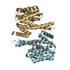
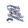
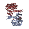
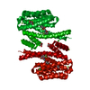
| Entry | Database: PDB / ID: 6yr5 | ||||||
|---|---|---|---|---|---|---|---|
| Title | 14-3-3 sigma in complex with hDMX-367 peptide | ||||||
 Components Components |
| ||||||
 Keywords Keywords | PEPTIDE BINDING PROTEIN | ||||||
| Function / homology |  Function and homology information Function and homology informationatrial septum development / heart valve development / atrioventricular valve morphogenesis / ventricular septum development / endocardial cushion morphogenesis / regulation of epidermal cell division /  protein kinase C inhibitor activity / positive regulation of epidermal cell differentiation / keratinocyte development / keratinization ...atrial septum development / heart valve development / atrioventricular valve morphogenesis / ventricular septum development / endocardial cushion morphogenesis / regulation of epidermal cell division / protein kinase C inhibitor activity / positive regulation of epidermal cell differentiation / keratinocyte development / keratinization / Regulation of localization of FOXO transcription factors / keratinocyte proliferation / DNA damage response, signal transduction by p53 class mediator / phosphoserine residue binding / Activation of BAD and translocation to mitochondria / negative regulation of keratinocyte proliferation / establishment of skin barrier / SARS-CoV-2 targets host intracellular signalling and regulatory pathways / Chk1/Chk2(Cds1) mediated inactivation of Cyclin B:Cdk1 complex / protein kinase A signaling / negative regulation of stem cell proliferation / SARS-CoV-1 targets host intracellular signalling and regulatory pathways / RHO GTPases activate PKNs / transcription repressor complex / protein export from nucleus / negative regulation of innate immune response / protein sequestering activity / TP53 Regulates Transcription of Genes Involved in G2 Cell Cycle Arrest / release of cytochrome c from mitochondria / positive regulation of protein export from nucleus / stem cell proliferation / Translocation of SLC2A4 (GLUT4) to the plasma membrane / TP53 Regulates Metabolic Genes / Stabilization of p53 / negative regulation of protein kinase activity / negative regulation of cysteine-type endopeptidase activity involved in apoptotic process / Oncogene Induced Senescence / Regulation of TP53 Activity through Methylation / negative regulation of protein catabolic process / ubiquitin-protein transferase activity / intrinsic apoptotic signaling pathway in response to DNA damage / Regulation of TP53 Degradation / cellular response to hypoxia / positive regulation of cell growth / protein-containing complex assembly / Oxidative Stress Induced Senescence / Regulation of TP53 Activity through Phosphorylation / protein stabilization / protein ubiquitination / regulation of cell cycle / Ub-specific processing proteases / cadherin binding / negative regulation of cell population proliferation / negative regulation of DNA-templated transcription / negative regulation of apoptotic process / protein kinase binding / negative regulation of transcription by RNA polymerase II / enzyme binding / signal transduction / extracellular space / extracellular exosome / zinc ion binding / nucleoplasm / identical protein binding / nucleus / cytosol / cytoplasm protein kinase C inhibitor activity / positive regulation of epidermal cell differentiation / keratinocyte development / keratinization ...atrial septum development / heart valve development / atrioventricular valve morphogenesis / ventricular septum development / endocardial cushion morphogenesis / regulation of epidermal cell division / protein kinase C inhibitor activity / positive regulation of epidermal cell differentiation / keratinocyte development / keratinization / Regulation of localization of FOXO transcription factors / keratinocyte proliferation / DNA damage response, signal transduction by p53 class mediator / phosphoserine residue binding / Activation of BAD and translocation to mitochondria / negative regulation of keratinocyte proliferation / establishment of skin barrier / SARS-CoV-2 targets host intracellular signalling and regulatory pathways / Chk1/Chk2(Cds1) mediated inactivation of Cyclin B:Cdk1 complex / protein kinase A signaling / negative regulation of stem cell proliferation / SARS-CoV-1 targets host intracellular signalling and regulatory pathways / RHO GTPases activate PKNs / transcription repressor complex / protein export from nucleus / negative regulation of innate immune response / protein sequestering activity / TP53 Regulates Transcription of Genes Involved in G2 Cell Cycle Arrest / release of cytochrome c from mitochondria / positive regulation of protein export from nucleus / stem cell proliferation / Translocation of SLC2A4 (GLUT4) to the plasma membrane / TP53 Regulates Metabolic Genes / Stabilization of p53 / negative regulation of protein kinase activity / negative regulation of cysteine-type endopeptidase activity involved in apoptotic process / Oncogene Induced Senescence / Regulation of TP53 Activity through Methylation / negative regulation of protein catabolic process / ubiquitin-protein transferase activity / intrinsic apoptotic signaling pathway in response to DNA damage / Regulation of TP53 Degradation / cellular response to hypoxia / positive regulation of cell growth / protein-containing complex assembly / Oxidative Stress Induced Senescence / Regulation of TP53 Activity through Phosphorylation / protein stabilization / protein ubiquitination / regulation of cell cycle / Ub-specific processing proteases / cadherin binding / negative regulation of cell population proliferation / negative regulation of DNA-templated transcription / negative regulation of apoptotic process / protein kinase binding / negative regulation of transcription by RNA polymerase II / enzyme binding / signal transduction / extracellular space / extracellular exosome / zinc ion binding / nucleoplasm / identical protein binding / nucleus / cytosol / cytoplasmSimilarity search - Function | ||||||
| Biological species |  Homo sapiens (human) Homo sapiens (human) | ||||||
| Method | X-RAY DIFFRACTION / SYNCHROTRON / MOLECULAR REPLACEMENT / Resolution: 2.25 Å | ||||||
 Authors Authors | Wolter, M. / Srdanovic, S. / Ottman, C. / Warriner, S. / Wilson, A. | ||||||
| Funding support | European Union, 1items
| ||||||
 Citation Citation | Journal: Febs J. / Year: 2022 Title: Understanding the interaction of 14-3-3 proteins with hDMX and hDM2: a structural and biophysical study. Authors: Srdanovic, S. / Wolter, M. / Trinh, C.H. / Ottmann, C. / Warriner, S.L. / Wilson, A.J. | ||||||
| History |
|
- Structure visualization
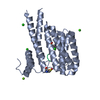
| Structure viewer | Molecule: MolmilJmol/JSmol |
|---|
- Downloads & links
-Download
| PDBx/mmCIF format | 6yr5.cif.gz | 198.5 KB | Display | PDBx/mmCIF format |
|---|---|---|---|---|
| PDB format | pdb6yr5.ent.gz | 157.4 KB | Display | PDB format |
| PDBx/mmJSON format | 6yr5.json.gz | Tree view | PDBx/mmJSON format | |
| Others |  Other downloads Other downloads |
-Validation report
| Arichive directory | https://data.pdbj.org/pub/pdb/validation_reports/yr/6yr5ftp://data.pdbj.org/pub/pdb/validation_reports/yr/6yr5 | HTTPS FTP |
|---|
-Related structure data
| Related structure data | 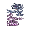 6yr6C 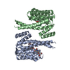 6yr7C  4datS C: citing same article ( S: Starting model for refinement |
|---|---|
| Similar structure data |


-Links
 PDBj
PDBj


- Assembly
| Deposited unit | 
| ||||||||
|---|---|---|---|---|---|---|---|---|---|
| 1 | 
| ||||||||
| 2 | 
| ||||||||
| Unit cell |
|
-Components
| #1: Protein | Mass: 26542.914 Da / Num. of mol.: 4 Source method: isolated from a genetically manipulated source Source: (gene. exp.) Homo sapiens (human) / Gene: SFN, HME1 / Production host:  Escherichia coli (E. coli) / References: UniProt: P31947 Escherichia coli (E. coli) / References: UniProt: P31947#2: Protein/peptide | Mass: 1681.894 Da / Num. of mol.: 4 / Source method: obtained synthetically / Source: (synth.) Homo sapiens (human) / References: UniProt: O15151#3: Chemical | ChemComp-SO4 / | Sulfate  Mass: 96.063 Da / Num. of mol.: 1 / Source method: obtained synthetically / Formula: SO4 Mass: 96.063 Da / Num. of mol.: 1 / Source method: obtained synthetically / Formula: SO4#4: Water | ChemComp-HOH / | Water Mass: 18.015 Da / Num. of mol.: 371 / Source method: isolated from a natural source / Formula: H2O Mass: 18.015 Da / Num. of mol.: 371 / Source method: isolated from a natural source / Formula: H2OHas ligand of interest | N | |
|---|
-Experimental details
-Experiment
| Experiment | Method: X-RAY DIFFRACTION / Number of used crystals: 1 |
|---|
- Sample preparation
| Crystal | Density Matthews: 3.04 Å3/Da / Density % sol: 59.58 % |
|---|---|
| Crystal grow | Temperature: 277 K / Method: vapor diffusion, hanging drop / pH: 7 Details: 0.1M Bis-Tris propane pH 7, 0.2 M Sodium citrate, 20% PEG3350 |
-Data collection
| Diffraction | Mean temperature: 100 K / Serial crystal experiment: N | ||||||||||||||||||||||||||||||
|---|---|---|---|---|---|---|---|---|---|---|---|---|---|---|---|---|---|---|---|---|---|---|---|---|---|---|---|---|---|---|---|
| Diffraction source | Source: SYNCHROTRON / Site: PETRA III, DESY  / Beamline: P11 / Wavelength: 1.033208 Å / Beamline: P11 / Wavelength: 1.033208 Å | ||||||||||||||||||||||||||||||
| Detector | Type: DECTRIS PILATUS 6M-F / Detector: PIXEL / Date: Apr 5, 2018 | ||||||||||||||||||||||||||||||
| Radiation | Protocol: SINGLE WAVELENGTH / Monochromatic (M) / Laue (L): M / Scattering type: x-ray | ||||||||||||||||||||||||||||||
| Radiation wavelength | Wavelength: 1.033208 Å / Relative weight: 1 | ||||||||||||||||||||||||||||||
| Reflection | Resolution: 2.1→74.53 Å / Num. obs: 75667 / % possible obs: 97.4 % / Redundancy: 5.8 % / CC1/2: 0.99 / Rmerge(I) obs: 0.114 / Rpim(I) all: 0.051 / Rrim(I) all: 0.125 / Net I/σ(I): 9.5 | ||||||||||||||||||||||||||||||
| Reflection shell | Diffraction-ID: 1
|
- Processing
| Software |
| ||||||||||||||||||||||||||||||||||||||||||||||||||||||||||||
|---|---|---|---|---|---|---|---|---|---|---|---|---|---|---|---|---|---|---|---|---|---|---|---|---|---|---|---|---|---|---|---|---|---|---|---|---|---|---|---|---|---|---|---|---|---|---|---|---|---|---|---|---|---|---|---|---|---|---|---|---|---|
| Refinement | Method to determine structure: MOLECULAR REPLACEMENT Starting model: 4DAT Resolution: 2.25→74.5 Å / Cor.coef. Fo:Fc: 0.93 / Cor.coef. Fo:Fc free: 0.92 / SU B: 5.156 / SU ML: 0.128 / Cross valid method: FREE R-VALUE / σ(F): 0 / ESU R: 0.24 / ESU R Free: 0.186 Details: HYDROGENS HAVE BEEN ADDED IN THE RIDING POSITIONS U VALUES : REFINED INDIVIDUALLY
| ||||||||||||||||||||||||||||||||||||||||||||||||||||||||||||
| Solvent computation | Ion probe radii: 0.8 Å / Shrinkage radii: 0.8 Å / VDW probe radii: 1.2 Å | ||||||||||||||||||||||||||||||||||||||||||||||||||||||||||||
| Displacement parameters | Biso max: 105.17 Å2 / Biso mean: 31.365 Å2 / Biso min: 11.36 Å2
| ||||||||||||||||||||||||||||||||||||||||||||||||||||||||||||
| Refinement step | Cycle: final / Resolution: 2.25→74.5 Å
| ||||||||||||||||||||||||||||||||||||||||||||||||||||||||||||
| Refine LS restraints |
| ||||||||||||||||||||||||||||||||||||||||||||||||||||||||||||
| LS refinement shell | Resolution: 2.25→2.308 Å / Rfactor Rfree error: 0 / Total num. of bins used: 20
|
