 Movie
Movie Controller


+ Open data

- Basic information
| Entry | Database: PDB / ID: 6xrc | ||||||
|---|---|---|---|---|---|---|---|
| Title | Apo NIS synthetase DesD variant R306Q | ||||||
 Components Components | Desferrioxamine E biosynthesis protein DesD | ||||||
 Keywords Keywords |  BIOSYNTHETIC PROTEIN / NIS synthetase / catalytic variant / siderophore synthesis BIOSYNTHETIC PROTEIN / NIS synthetase / catalytic variant / siderophore synthesis | ||||||
| Function / homology | Aerobactin siderophore biosynthesis, IucA/IucC, N-terminal / Aerobactin siderophore biosynthesis, IucA/IucC-like / IucA / IucC family / Ferric iron reductase FhuF domain / Ferric iron reductase FhuF-like transporter / acid-amino acid ligase activity / siderophore biosynthetic process / ADENOSINE-5'-TRIPHOSPHATE / Uncharacterized protein Function and homology information Function and homology information | ||||||
| Biological species |  Streptomyces coelicolor (bacteria) Streptomyces coelicolor (bacteria) | ||||||
| Method | X-RAY DIFFRACTION / SYNCHROTRON / MOLECULAR REPLACEMENT / Resolution: 2.45 Å | ||||||
 Authors Authors | Hoffmann, K.M. | ||||||
| Funding support |  United States, 1items United States, 1items
| ||||||
 Citation Citation | Journal: Biochemistry / Year: 2020 Title: Cofactor Complexes of DesD, a Model Enzyme in the Virulence-related NIS Synthetase Family. Authors: Hoffmann, K.M. / Goncuian, E.S. / Karimi, K.L. / Amendola, C.R. / Mojab, Y. / Wood, K.M. / Prussia, G.A. / Nix, J. / Yamamoto, M. / Lathan, K. / Orion, I.W. | ||||||
| History |
|
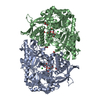
- Structure visualization
| Structure viewer | Molecule: MolmilJmol/JSmol |
|---|
- Downloads & links
-Download
| PDBx/mmCIF format | 6xrc.cif.gz | 815.7 KB | Display | PDBx/mmCIF format |
|---|---|---|---|---|
| PDB format | pdb6xrc.ent.gz | 673.6 KB | Display | PDB format |
| PDBx/mmJSON format | 6xrc.json.gz | Tree view | PDBx/mmJSON format | |
| Others |  Other downloads Other downloads |
-Validation report
| Arichive directory | https://data.pdbj.org/pub/pdb/validation_reports/xr/6xrcftp://data.pdbj.org/pub/pdb/validation_reports/xr/6xrc | HTTPS FTP |
|---|
-Related structure data
| Related structure data | 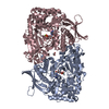 6p63C  6nl2S S: Starting model for refinement C: citing same article ( |
|---|---|
| Similar structure data |


-Links
 PDBj
PDBj- Assembly
| Deposited unit | 
| ||||||||
|---|---|---|---|---|---|---|---|---|---|
| 1 |
| ||||||||
| Unit cell |
|
-Components
| #1: Protein | Mass: 67706.320 Da / Num. of mol.: 2 / Mutation: R306Q Source method: isolated from a genetically manipulated source Source: (gene. exp.) Streptomyces coelicolor (bacteria) / Gene: SCO2785 / Production host: Escherichia coli (E. coli) / References: UniProt: Q9L069#2: Chemical | Glycerol  Mass: 92.094 Da / Num. of mol.: 3 / Source method: obtained synthetically / Formula: C3H8O3 Mass: 92.094 Da / Num. of mol.: 3 / Source method: obtained synthetically / Formula: C3H8O3#3: Chemical | Adenosine triphosphate  Mass: 507.181 Da / Num. of mol.: 2 / Source method: obtained synthetically / Formula: C10H16N5O13P3 / Comment: ATP, energy-carrying molecule*YM Mass: 507.181 Da / Num. of mol.: 2 / Source method: obtained synthetically / Formula: C10H16N5O13P3 / Comment: ATP, energy-carrying molecule*YM#4: Chemical |   Mass: 24.305 Da / Num. of mol.: 3 / Source method: obtained synthetically / Formula: Mg Mass: 24.305 Da / Num. of mol.: 3 / Source method: obtained synthetically / Formula: Mg#5: Water | ChemComp-HOH / | Water Mass: 18.015 Da / Num. of mol.: 640 / Source method: isolated from a natural source / Formula: H2O Mass: 18.015 Da / Num. of mol.: 640 / Source method: isolated from a natural source / Formula: H2OHas ligand of interest | Y | |
|---|
-Experimental details
-Experiment
| Experiment | Method: X-RAY DIFFRACTION / Number of used crystals: 1 |
|---|
- Sample preparation
| Crystal | Density Matthews: 2.54 Å3/Da / Density % sol: 51.66 % |
|---|---|
| Crystal grow | Temperature: 298 K / Method: vapor diffusion, hanging drop / pH: 8.5 Details: 0.08 mM Tris-HCl, pH 8.5, 160 mM magnesium chloride, 24% PEG4000, 20% glycerol |
-Data collection
| Diffraction | Mean temperature: 80 K / Serial crystal experiment: N |
|---|---|
| Diffraction source | Source: SYNCHROTRON / Site: ALS / Beamline: 4.2.2 / Wavelength: 1 Å |
| Detector | Type: RDI CMOS_8M / Detector: CMOS / Date: Nov 30, 2016 |
| Radiation | Monochromator: double crystal Si(111) / Protocol: SINGLE WAVELENGTH / Monochromatic (M) / Laue (L): M / Scattering type: x-ray |
| Radiation wavelength | Wavelength: 1 Å / Relative weight: 1 |
| Reflection | Resolution: 2.45→61.12 Å / Num. obs: 50570 / % possible obs: 99.9 % / Redundancy: 7.4 % / CC1/2: 0.993 / Rmerge(I) obs: 0.153 / Rpim(I) all: 0.063 / Rrim(I) all: 0.172 / Χ2: 0.98 / Net I/σ(I): 7.7 |
| Reflection shell | Resolution: 2.45→2.53 Å / Rmerge(I) obs: 0.534 / Mean I/σ(I) obs: 3.4 / Num. unique obs: 4574 / CC1/2: 0.964 / Χ2: 0.74 |
- Processing
| Software |
| ||||||||||||||||||||||||||||||||||||||||||||||||||||||||||||||||||||||||||||||||||||||||||||||||||||||||||||||||||||||||||||||||||||||||||||||||||||||||||||||||||||||||||||||||||||||
|---|---|---|---|---|---|---|---|---|---|---|---|---|---|---|---|---|---|---|---|---|---|---|---|---|---|---|---|---|---|---|---|---|---|---|---|---|---|---|---|---|---|---|---|---|---|---|---|---|---|---|---|---|---|---|---|---|---|---|---|---|---|---|---|---|---|---|---|---|---|---|---|---|---|---|---|---|---|---|---|---|---|---|---|---|---|---|---|---|---|---|---|---|---|---|---|---|---|---|---|---|---|---|---|---|---|---|---|---|---|---|---|---|---|---|---|---|---|---|---|---|---|---|---|---|---|---|---|---|---|---|---|---|---|---|---|---|---|---|---|---|---|---|---|---|---|---|---|---|---|---|---|---|---|---|---|---|---|---|---|---|---|---|---|---|---|---|---|---|---|---|---|---|---|---|---|---|---|---|---|---|---|---|---|
| Refinement | Method to determine structure: MOLECULAR REPLACEMENT Starting model: PDB entry 6NL2 Resolution: 2.45→59.47 Å / Cor.coef. Fo:Fc: 0.947 / Cor.coef. Fo:Fc free: 0.909 / Cross valid method: FREE R-VALUE / ESU R: 0.207 / ESU R Free: 0.099 Details: HYDROGENS HAVE BEEN ADDED IN THEIR RIDING POSITIONS
| ||||||||||||||||||||||||||||||||||||||||||||||||||||||||||||||||||||||||||||||||||||||||||||||||||||||||||||||||||||||||||||||||||||||||||||||||||||||||||||||||||||||||||||||||||||||
| Solvent computation | Ion probe radii: 0.8 Å / Shrinkage radii: 0.8 Å / VDW probe radii: 1.2 Å / Solvent model: MASK BULK SOLVENT | ||||||||||||||||||||||||||||||||||||||||||||||||||||||||||||||||||||||||||||||||||||||||||||||||||||||||||||||||||||||||||||||||||||||||||||||||||||||||||||||||||||||||||||||||||||||
| Displacement parameters | Biso mean: 46.18 Å2
| ||||||||||||||||||||||||||||||||||||||||||||||||||||||||||||||||||||||||||||||||||||||||||||||||||||||||||||||||||||||||||||||||||||||||||||||||||||||||||||||||||||||||||||||||||||||
| Refinement step | Cycle: LAST / Resolution: 2.45→59.47 Å
| ||||||||||||||||||||||||||||||||||||||||||||||||||||||||||||||||||||||||||||||||||||||||||||||||||||||||||||||||||||||||||||||||||||||||||||||||||||||||||||||||||||||||||||||||||||||
| Refine LS restraints |
|