[English] 日本語
Yorodumi
- PDB-5o7o: The crystal structure of DfoC, the desferrioxamine biosynthetic p... -

+
Open data


ID or keywords:

Loading...

-
Basic information

Entry
Database: PDB / ID: 5o7o
Title
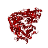
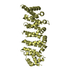
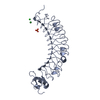
The crystal structure of DfoC, the desferrioxamine biosynthetic pathway acetyltransferase/Non-Ribosomal Peptide Synthetase (NRPS)-Independent Siderophore (NIS) from the fire blight disease pathogen Erwinia amylovora
ComponentsDesferrioxamine siderophore biosynthesis protein dfoC
KeywordsBIOSYNTHETIC PROTEIN / siderophore biosynthesis / desferrioxamine / fire blight
Function / homology
Function and homology information


siderophore biosynthetic process / acyltransferase activity
Similarity search - Function
Acyltransferase MbtK/IucB-like, conserved domain / Siderophore biosynthesis protein domain / Transferase(Phosphotransferase); domain 1 - #40 / TATA-Binding Protein - #280 / Single alpha-helices involved in coiled-coils or other helix-helix interfaces - #3370 / Aerobactin siderophore biosynthesis, IucA/IucC, N-terminal / Aerobactin siderophore biosynthesis, IucA/IucC-like / IucA / IucC family / Ferric iron reductase FhuF domain / Ferric iron reductase FhuF-like transporter ...Acyltransferase MbtK/IucB-like, conserved domain / Siderophore biosynthesis protein domain / Transferase(Phosphotransferase); domain 1 - #40 / TATA-Binding Protein - #280 / Single alpha-helices involved in coiled-coils or other helix-helix interfaces - #3370 / Aerobactin siderophore biosynthesis, IucA/IucC, N-terminal / Aerobactin siderophore biosynthesis, IucA/IucC-like / IucA / IucC family / Ferric iron reductase FhuF domain / Ferric iron reductase FhuF-like transporter / Acetyltransferase (GNAT) domain / TATA-Binding Protein / Single alpha-helices involved in coiled-coils or other helix-helix interfaces / Acyl-CoA N-acyltransferase / Helix non-globular / Special / Transferase(Phosphotransferase); domain 1 / 2-Layer Sandwich / Orthogonal Bundle / Mainly Alpha / Alpha Beta
Similarity search - Domain/homology
Desferrioxamine siderophore biosynthesis protein dfoC
Similarity search - Component
Biological speciesErwinia amylovora CFBP1430 (bacteria)
MethodX-RAY DIFFRACTION / SYNCHROTRON / MOLECULAR REPLACEMENT / Resolution: 2.11 Å
AuthorsSalomone-Stagni, M. / Bartho, J.D. / Polsinelli, I. / Bellini, D. / Walsh, M.A. / Demitri, N. / Benini, S.
Funding support Italy, 2items
OrganizationGrant numberCountry
Autonomous Province of Bolzano Italy
Free University of Bolzano Italy
CitationJournal: J. Struct. Biol. / Year: 2018
Title: A complete structural characterization of the desferrioxamine E biosynthetic pathway from the fire blight pathogen Erwinia amylovora.
Authors: Salomone-Stagni, M. / Bartho, J.D. / Polsinelli, I. / Bellini, D. / Walsh, M.A. / Demitri, N. / Benini, S.
History
DepositionJun 9, 2017Deposition site: PDBE / Processing site: PDBE
Revision 1.0Feb 28, 2018Provider: repository / Type: Initial release
Revision 1.1May 23, 2018Group: Data collection / Database references / Category: citation
Item: _citation.journal_volume / _citation.page_first / _citation.page_last
Revision 1.2Jan 17, 2024Group: Data collection / Database references / Refinement description
Category: chem_comp_atom / chem_comp_bond ...chem_comp_atom / chem_comp_bond / database_2 / pdbx_initial_refinement_model
Item: _database_2.pdbx_DOI / _database_2.pdbx_database_accession

-
Structure visualization

Structure viewerMolecule:
MolmilJmol/JSmol

Downloads & links

-
Assembly

Deposited unit
A: Desferrioxamine siderophore biosynthesis protein dfoC
B: Desferrioxamine siderophore biosynthesis protein dfoC
C: Desferrioxamine siderophore biosynthesis protein dfoC
D: Desferrioxamine siderophore biosynthesis protein dfoC


Theoretical massNumber of molelcules
Total (without water)359,6284
Polymers359,6284
Non-polymers00
Water7,656425
1
A: Desferrioxamine siderophore biosynthesis protein dfoC
B: Desferrioxamine siderophore biosynthesis protein dfoC


Theoretical massNumber of molelcules
Total (without water)179,8142
Polymers179,8142
Non-polymers00
Water362
TypeNameSymmetry operationNumber
identity operation1_555x,y,z1
MethodPISA
2
C: Desferrioxamine siderophore biosynthesis protein dfoC
D: Desferrioxamine siderophore biosynthesis protein dfoC


Theoretical massNumber of molelcules
Total (without water)179,8142
Polymers179,8142
Non-polymers00
Water362
TypeNameSymmetry operationNumber
identity operation1_555x,y,z1
MethodPISA
Unit cell
Length a, b, c (Å)75.170, 156.350, 93.810
Angle α, β, γ (deg.)90.00, 95.57, 90.00
Int Tables number4
Space group name H-MP1211

-
Components

#1: Protein
Desferrioxamine siderophore biosynthesis protein dfoC


Mass: 89907.023 Da / Num. of mol.: 4
Source method: isolated from a genetically manipulated source
Details: DfoC N-terminal domain / Source: (gene. exp.) Erwinia amylovora CFBP1430 (bacteria) / Gene: dfoC, EAMY_3240 / Production host: Escherichia coli (E. coli) / References: UniProt: D4I247
#2: Water ChemComp-HOH / water / Water


Mass: 18.015 Da / Num. of mol.: 425 / Source method: isolated from a natural source / Formula: H2O

-
Experimental details

-
Experiment

ExperimentMethod: X-RAY DIFFRACTION / Number of used crystals: 1

-
Sample preparation

Crystal growTemperature: 293 K / Method: vapor diffusion / pH: 7
Details: 0.1 M TRIS-HCl, 0.2 M MgCl2, 0.06 M Ammonium citrate, 10% PEG 8K

-
Data collection

DiffractionMean temperature: 100 K
Diffraction sourceSource: SYNCHROTRON / Site: Diamond / Beamline: I04 / Wavelength: 0.97625 Å
DetectorType: DECTRIS PILATUS 6M / Detector: PIXEL / Date: Jul 16, 2015
RadiationProtocol: SINGLE WAVELENGTH / Monochromatic (M) / Laue (L): M / Scattering type: x-ray
Radiation wavelengthWavelength: 0.97625 Å / Relative weight: 1
ReflectionResolution: 2.11→78.18 Å / Num. obs: 123387 / % possible obs: 99.79 % / Redundancy: 3.4 % / Rrim(I) all: 0.04 / Net I/σ(I): 12
Reflection shellResolution: 2.11→2.165 Å / Num. unique obs: 9132

-
Processing

Software
NameVersionClassification
REFMAC5.8.0158refinement
XDSdata reduction
XSCALEdata scaling
MOLREPphasing
RefinementMethod to determine structure: MOLECULAR REPLACEMENT
Starting model: 2x0o
Resolution: 2.11→78.18 Å / Cor.coef. Fo:Fc: 0.952 / Cor.coef. Fo:Fc free: 0.932 / SU B: 8.851 / SU ML: 0.207 / Cross valid method: THROUGHOUT / ESU R: 0.197 / ESU R Free: 0.181 / Details: HYDROGENS HAVE BEEN ADDED IN THE RIDING POSITIONS
RfactorNum. reflection% reflectionSelection details
Rfree0.264 6119 5 %RANDOM
Rwork0.22055 ---
obs0.2227 117229 99.79 %-
Solvent computationIon probe radii: 0.8 Å / Shrinkage radii: 0.8 Å / VDW probe radii: 1.2 Å
Displacement parametersBiso mean: 50.695 Å2
Baniso -1Baniso -2Baniso -3
1--0.19 Å20 Å2-4.14 Å2
2---3.38 Å2-0 Å2
3---4.29 Å2
Refinement stepCycle: 1 / Resolution: 2.11→78.18 Å
ProteinNucleic acidLigandSolventTotal
Num. atoms11584 0 0 425 12009
Refine LS restraints
Refine-IDTypeDev idealDev ideal targetNumber
X-RAY DIFFRACTIONr_bond_refined_d0.0160.01911888
X-RAY DIFFRACTIONr_bond_other_d0.0030.0210737
X-RAY DIFFRACTIONr_angle_refined_deg1.6791.93516147
X-RAY DIFFRACTIONr_angle_other_deg1.04324857
X-RAY DIFFRACTIONr_dihedral_angle_1_deg6.85351433
X-RAY DIFFRACTIONr_dihedral_angle_2_deg36.28623.802597
X-RAY DIFFRACTIONr_dihedral_angle_3_deg14.775151937
X-RAY DIFFRACTIONr_dihedral_angle_4_deg17.8731578
X-RAY DIFFRACTIONr_chiral_restr0.0980.21745
X-RAY DIFFRACTIONr_gen_planes_refined0.0080.02113295
X-RAY DIFFRACTIONr_gen_planes_other0.0020.022563
X-RAY DIFFRACTIONr_nbd_refined
X-RAY DIFFRACTIONr_nbd_other
X-RAY DIFFRACTIONr_nbtor_refined
X-RAY DIFFRACTIONr_nbtor_other
X-RAY DIFFRACTIONr_xyhbond_nbd_refined
X-RAY DIFFRACTIONr_xyhbond_nbd_other
X-RAY DIFFRACTIONr_metal_ion_refined
X-RAY DIFFRACTIONr_metal_ion_other
X-RAY DIFFRACTIONr_symmetry_vdw_refined
X-RAY DIFFRACTIONr_symmetry_vdw_other
X-RAY DIFFRACTIONr_symmetry_hbond_refined
X-RAY DIFFRACTIONr_symmetry_hbond_other
X-RAY DIFFRACTIONr_symmetry_metal_ion_refined
X-RAY DIFFRACTIONr_symmetry_metal_ion_other
X-RAY DIFFRACTIONr_mcbond_it3.1895.0575756
X-RAY DIFFRACTIONr_mcbond_other3.1895.0575755
X-RAY DIFFRACTIONr_mcangle_it4.4337.5717181
X-RAY DIFFRACTIONr_mcangle_other4.4327.5717182
X-RAY DIFFRACTIONr_scbond_it3.4315.2756132
X-RAY DIFFRACTIONr_scbond_other3.4315.2766133
X-RAY DIFFRACTIONr_scangle_it
X-RAY DIFFRACTIONr_scangle_other4.8987.8138967
X-RAY DIFFRACTIONr_long_range_B_refined6.65958.40513227
X-RAY DIFFRACTIONr_long_range_B_other6.64358.33713184
X-RAY DIFFRACTIONr_rigid_bond_restr
X-RAY DIFFRACTIONr_sphericity_free
X-RAY DIFFRACTIONr_sphericity_bonded
LS refinement shellResolution: 2.11→2.165 Å
RfactorNum. reflection% reflection
Rfree0.34 462 5 %
Rwork0.35 8668 -
obs--99.85 %

+
About Yorodumi

-
News

-
Feb 9, 2022. New format data for meta-information of EMDB entries

New format data for meta-information of EMDB entries

  • Version 3 of the EMDB header file is now the official format.
  • The previous official version 1.9 will be removed from the archive.

Related info.:EMDB header

External links:wwPDB to switch to version 3 of the EMDB data model

-
Aug 12, 2020. Covid-19 info

Covid-19 info

URL: https://pdbj.org/emnavi/covid19.php

New page: Covid-19 featured information page in EM Navigator.

Related info.:Covid-19 info / Mar 5, 2020. Novel coronavirus structure data

+
Mar 5, 2020. Novel coronavirus structure data

Novel coronavirus structure data

Related info.:Yorodumi Speices / Aug 12, 2020. Covid-19 info

External links:COVID-19 featured content - PDBj / Molecule of the Month (242):Coronavirus Proteases

+
Jan 31, 2019. EMDB accession codes are about to change! (news from PDBe EMDB page)

EMDB accession codes are about to change! (news from PDBe EMDB page)

  • The allocation of 4 digits for EMDB accession codes will soon come to an end. Whilst these codes will remain in use, new EMDB accession codes will include an additional digit and will expand incrementally as the available range of codes is exhausted. The current 4-digit format prefixed with “EMD-” (i.e. EMD-XXXX) will advance to a 5-digit format (i.e. EMD-XXXXX), and so on. It is currently estimated that the 4-digit codes will be depleted around Spring 2019, at which point the 5-digit format will come into force.
  • The EM Navigator/Yorodumi systems omit the EMD- prefix.

Related info.:Q: What is EMD? / ID/Accession-code notation in Yorodumi/EM Navigator

External links:EMDB Accession Codes are Changing Soon! / Contact to PDBj

+
Jul 12, 2017. Major update of PDB

Major update of PDB

  • wwPDB released updated PDB data conforming to the new PDBx/mmCIF dictionary.
  • This is a major update changing the version number from 4 to 5, and with Remediation, in which all the entries are updated.
  • In this update, many items about electron microscopy experimental information are reorganized (e.g. em_software).
  • Now, EM Navigator and Yorodumi are based on the updated data.

External links:wwPDB Remediation / Enriched Model Files Conforming to OneDep Data Standards Now Available in the PDB FTP Archive

-
Yorodumi

Thousand views of thousand structures

  • Yorodumi is a browser for structure data from EMDB, PDB, SASBDB, etc.
  • This page is also the successor to EM Navigator detail page, and also detail information page/front-end page for Omokage search.
  • The word "yorodu" (or yorozu) is an old Japanese word meaning "ten thousand". "mi" (miru) is to see.

Related info.:EMDB / PDB / SASBDB / Comparison of 3 databanks / Yorodumi Search / Aug 31, 2016. New EM Navigator & Yorodumi / Yorodumi Papers / Jmol/JSmol / Function and homology information / Changes in new EM Navigator and Yorodumi

Read more