 Movie
Movie Controller

[English] 日本語
 Yorodumi
- PDB-6x8p: Crystal structure of 3D11 Fab in complex with Plasmodium berghei ... -
+ Open data

- Basic information
| Entry | Database: PDB / ID: 6x8p | ||||||||||||||||||
|---|---|---|---|---|---|---|---|---|---|---|---|---|---|---|---|---|---|---|---|
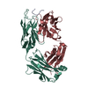
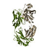
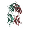
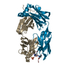
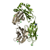
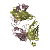
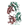
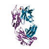
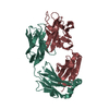
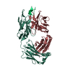
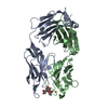
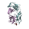
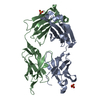
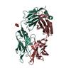
| Title | Crystal structure of 3D11 Fab in complex with Plasmodium berghei circumsporozoite protein NPND peptide | ||||||||||||||||||
 Components Components |
| ||||||||||||||||||
 Keywords Keywords |  IMMUNE SYSTEM / Malaria / Antibody IMMUNE SYSTEM / Malaria / Antibody | ||||||||||||||||||
| Function / homology |  Function and homology information Function and homology informationpositive regulation by symbiont of entry into host / positive regulation of development of symbiont in host / positive regulation of developmental process / adhesion of symbiont to host cell / entry into host cell by a symbiont-containing vacuole / external side of plasma membrane / cytoplasmSimilarity search - Function | ||||||||||||||||||
| Biological species |  Mus musculus (house mouse) Mus musculus (house mouse) Plasmodium berghei ANKA (eukaryote) Plasmodium berghei ANKA (eukaryote) | ||||||||||||||||||
| Method | X-RAY DIFFRACTION / SYNCHROTRON / MOLECULAR REPLACEMENT / Resolution: 2.27 Å | ||||||||||||||||||
 Authors Authors | Thai, E. / Julien, J.P. | ||||||||||||||||||
| Funding support |  United States, United States,  Canada, 5items Canada, 5items
| ||||||||||||||||||
 Citation Citation | Journal: Elife / Year: 2020 Title: Structural ordering of the circumsporozoite protein repeats by inhibitory antibody 3D11. Authors: Iga Kucharska / Elaine Thai / Ananya Srivastava / John L Rubinstein / Régis Pomès / Jean-Philippe Julien / Abstract: Plasmodium sporozoites express circumsporozoite protein (CSP) on their surface, an essential protein that contains central repeating motifs. Antibodies targeting this region can neutralize infection, ...Plasmodium sporozoites express circumsporozoite protein (CSP) on their surface, an essential protein that contains central repeating motifs. Antibodies targeting this region can neutralize infection, and the partial efficacy of RTS,S/AS01 - the leading malaria vaccine against (Pf) - has been associated with the humoral response against the repeats. Although structural details of antibody recognition of PfCSP have recently emerged, the molecular basis of antibody-mediated inhibition of other Plasmodium species via CSP binding remains unclear. Here, we analyze the structure and molecular interactions of potent monoclonal antibody (mAb) 3D11 binding to CSP (PbCSP) using molecular dynamics simulations, X-ray crystallography, and cryoEM. We reveal that mAb 3D11 can accommodate all subtle variances of the PbCSP repeating motifs, and, upon binding, induces structural ordering of PbCSP through homotypic interactions. Together, our findings uncover common mechanisms of antibody evolution in mammals against the CSP repeats of Plasmodium sporozoites. | ||||||||||||||||||
| History |
|
- Structure visualization
| Structure viewer | Molecule: MolmilJmol/JSmol |
|---|
- Downloads & links
-Download
| PDBx/mmCIF format | 6x8p.cif.gz | 226.8 KB | Display | PDBx/mmCIF format |
|---|---|---|---|---|
| PDB format | pdb6x8p.ent.gz | 151.2 KB | Display | PDB format |
| PDBx/mmJSON format | 6x8p.json.gz | Tree view | PDBx/mmJSON format | |
| Others |  Other downloads Other downloads |
-Validation report
| Arichive directory | https://data.pdbj.org/pub/pdb/validation_reports/x8/6x8pftp://data.pdbj.org/pub/pdb/validation_reports/x8/6x8p | HTTPS FTP |
|---|
-Related structure data
| Related structure data |  6x87C 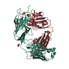 6x8qC 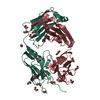 6x8sC 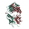 6x8uC 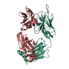 2eh7S S: Starting model for refinement C: citing same article ( |
|---|---|
| Similar structure data |


-Links
 PDBj
PDBj

- Assembly
| Deposited unit | 
| ||||||||||||
|---|---|---|---|---|---|---|---|---|---|---|---|---|---|
| 1 |
| ||||||||||||
| Unit cell |
| ||||||||||||
| Components on special symmetry positions |
|
-Components
| #1: Antibody | Mass: 22629.295 Da / Num. of mol.: 1 Source method: isolated from a genetically manipulated source Source: (gene. exp.) Mus musculus (house mouse) / Production host:  Homo sapiens (human) Homo sapiens (human) | ||||
|---|---|---|---|---|---|
| #2: Antibody | Mass: 23989.838 Da / Num. of mol.: 1 Source method: isolated from a genetically manipulated source Source: (gene. exp.) Mus musculus (house mouse) / Production host: Homo sapiens (human) | ||||
| #3: Protein/peptide | Mass: 1675.750 Da / Num. of mol.: 1 / Source method: obtained synthetically / Source: (synth.) Plasmodium berghei ANKA (eukaryote) / References: UniProt: P23093*PLUS | ||||
| #4: Chemical | ChemComp-EDO / Ethylene glycol  Mass: 62.068 Da / Num. of mol.: 6 / Source method: obtained synthetically / Formula: C2H6O2 Mass: 62.068 Da / Num. of mol.: 6 / Source method: obtained synthetically / Formula: C2H6O2#5: Water | ChemComp-HOH / | Water Mass: 18.015 Da / Num. of mol.: 259 / Source method: isolated from a natural source / Formula: H2O Mass: 18.015 Da / Num. of mol.: 259 / Source method: isolated from a natural source / Formula: H2OHas ligand of interest | N | |
-Experimental details
-Experiment
| Experiment | Method: X-RAY DIFFRACTION / Number of used crystals: 1 |
|---|
- Sample preparation
| Crystal | Density Matthews: 2.52 Å3/Da / Density % sol: 51.12 % |
|---|---|
| Crystal grow | Temperature: 293.15 K / Method: vapor diffusion Details: 25% (w/v) PEG 3350, 0.2 M lithium sulfate, 0.1 M Tris pH 8.5 |
-Data collection
| Diffraction | Mean temperature: 100 K / Serial crystal experiment: N |
|---|---|
| Diffraction source | Source: SYNCHROTRON / Site: NSLS-II / Beamline: 17-ID-1 / Wavelength: 0.979329 Å |
| Detector | Type: DECTRIS EIGER X 9M / Detector: PIXEL / Date: Feb 5, 2019 |
| Radiation | Protocol: SINGLE WAVELENGTH / Monochromatic (M) / Laue (L): M / Scattering type: x-ray |
| Radiation wavelength | Wavelength: 0.979329 Å / Relative weight: 1 |
| Reflection | Resolution: 2.27→40 Å / Num. obs: 23398 / % possible obs: 99.3 % / Redundancy: 19.1 % / Biso Wilson estimate: 31.96 Å2 / CC1/2: 0.999 / Net I/σ(I): 19 |
| Reflection shell | Resolution: 2.27→2.37 Å / Num. unique obs: 2556 / CC1/2: 0.935 / % possible all: 94.4 |
- Processing
| Software |
| ||||||||||||||||||||||||||||||||||||||||||||||||||||||||||||||||||||||||||||||||||||||||||||||||||||||||||||||||||||||||||||||||||||||||||||||||||||||
|---|---|---|---|---|---|---|---|---|---|---|---|---|---|---|---|---|---|---|---|---|---|---|---|---|---|---|---|---|---|---|---|---|---|---|---|---|---|---|---|---|---|---|---|---|---|---|---|---|---|---|---|---|---|---|---|---|---|---|---|---|---|---|---|---|---|---|---|---|---|---|---|---|---|---|---|---|---|---|---|---|---|---|---|---|---|---|---|---|---|---|---|---|---|---|---|---|---|---|---|---|---|---|---|---|---|---|---|---|---|---|---|---|---|---|---|---|---|---|---|---|---|---|---|---|---|---|---|---|---|---|---|---|---|---|---|---|---|---|---|---|---|---|---|---|---|---|---|---|---|---|---|
| Refinement | Method to determine structure: MOLECULAR REPLACEMENT Starting model: 2EH7 Resolution: 2.27→29.69 Å / SU ML: 0.2286 / Cross valid method: FREE R-VALUE / σ(F): 1.34 / Phase error: 20.9903 Stereochemistry target values: GeoStd + Monomer Library + CDL v1.2
| ||||||||||||||||||||||||||||||||||||||||||||||||||||||||||||||||||||||||||||||||||||||||||||||||||||||||||||||||||||||||||||||||||||||||||||||||||||||
| Solvent computation | Shrinkage radii: 0.9 Å / VDW probe radii: 1.11 Å / Solvent model: FLAT BULK SOLVENT MODEL | ||||||||||||||||||||||||||||||||||||||||||||||||||||||||||||||||||||||||||||||||||||||||||||||||||||||||||||||||||||||||||||||||||||||||||||||||||||||
| Displacement parameters | Biso mean: 35.16 Å2 | ||||||||||||||||||||||||||||||||||||||||||||||||||||||||||||||||||||||||||||||||||||||||||||||||||||||||||||||||||||||||||||||||||||||||||||||||||||||
| Refinement step | Cycle: LAST / Resolution: 2.27→29.69 Å
| ||||||||||||||||||||||||||||||||||||||||||||||||||||||||||||||||||||||||||||||||||||||||||||||||||||||||||||||||||||||||||||||||||||||||||||||||||||||
| Refine LS restraints |
| ||||||||||||||||||||||||||||||||||||||||||||||||||||||||||||||||||||||||||||||||||||||||||||||||||||||||||||||||||||||||||||||||||||||||||||||||||||||
| LS refinement shell |
| ||||||||||||||||||||||||||||||||||||||||||||||||||||||||||||||||||||||||||||||||||||||||||||||||||||||||||||||||||||||||||||||||||||||||||||||||||||||
| Refinement TLS params. | Method: refined / Refine-ID: X-RAY DIFFRACTION
| ||||||||||||||||||||||||||||||||||||||||||||||||||||||||||||||||||||||||||||||||||||||||||||||||||||||||||||||||||||||||||||||||||||||||||||||||||||||
| Refinement TLS group |
|
