[English] 日本語
Yorodumi
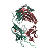
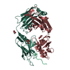
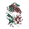
- PDB-6x8q: Crystal structure of 3D11 Fab in complex with Plasmodium berghei ... -

+
Open data


ID or keywords:

Loading...

-
Basic information

Entry
Database: PDB / ID: 6x8q
Title
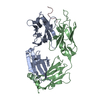
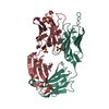
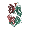
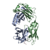
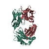
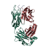
Crystal structure of 3D11 Fab in complex with Plasmodium berghei circumsporozoite protein PAPP peptide
Components
  • 3D11 Fab heavy chain
  • 3D11 Fab light chain
  • PAPP peptide
KeywordsIMMUNE SYSTEM / Malaria / Antibody
Function / homology
Function and homology information


positive regulation by symbiont of entry into host / positive regulation of development of symbiont in host / positive regulation of developmental process / adhesion of symbiont to host cell / entry into host cell by a symbiont-containing vacuole / external side of plasma membrane / cytoplasm
Similarity search - Function
Plasmodium circumsporozoite protein / Thrombospondin type 1 domain / Thrombospondin type-1 (TSP1) repeat superfamily / Thrombospondin type-1 (TSP1) repeat profile. / Thrombospondin type 1 repeats / Thrombospondin type-1 (TSP1) repeat
Similarity search - Domain/homology
Circumsporozoite protein
Similarity search - Component
Biological speciesMus musculus (house mouse)
Plasmodium berghei ANKA (eukaryote)
MethodX-RAY DIFFRACTION / SYNCHROTRON / MOLECULAR REPLACEMENT / Resolution: 1.6 Å
AuthorsThai, E. / Julien, J.P.
Funding support United States, Canada, 5items
OrganizationGrant numberCountry
National Institutes of Health/National Cancer Institute (NIH/NCI)ACB-12002 United States
National Institutes of Health/National Institute of General Medical Sciences (NIH/NIGMS)AGM-12006 United States
Other privateCIFAR Azrieli Global Scholar program Canada
Other governmentOntario Early Researcher Award program Canada
Other governmentCanada Research Chair program Canada
CitationJournal: Elife / Year: 2020
Title: Structural ordering of the circumsporozoite protein repeats by inhibitory antibody 3D11.
Authors: Iga Kucharska / Elaine Thai / Ananya Srivastava / John L Rubinstein / Régis Pomès / Jean-Philippe Julien /
Abstract: Plasmodium sporozoites express circumsporozoite protein (CSP) on their surface, an essential protein that contains central repeating motifs. Antibodies targeting this region can neutralize infection, ...Plasmodium sporozoites express circumsporozoite protein (CSP) on their surface, an essential protein that contains central repeating motifs. Antibodies targeting this region can neutralize infection, and the partial efficacy of RTS,S/AS01 - the leading malaria vaccine against (Pf) - has been associated with the humoral response against the repeats. Although structural details of antibody recognition of PfCSP have recently emerged, the molecular basis of antibody-mediated inhibition of other Plasmodium species via CSP binding remains unclear. Here, we analyze the structure and molecular interactions of potent monoclonal antibody (mAb) 3D11 binding to CSP (PbCSP) using molecular dynamics simulations, X-ray crystallography, and cryoEM. We reveal that mAb 3D11 can accommodate all subtle variances of the PbCSP repeating motifs, and, upon binding, induces structural ordering of PbCSP through homotypic interactions. Together, our findings uncover common mechanisms of antibody evolution in mammals against the CSP repeats of Plasmodium sporozoites.
History
DepositionJun 1, 2020Deposition site: RCSB / Processing site: RCSB
Revision 1.0Dec 2, 2020Provider: repository / Type: Initial release
Revision 1.1Dec 30, 2020Group: Database references / Category: citation / citation_author
Item: _citation.country / _citation.journal_abbrev ..._citation.country / _citation.journal_abbrev / _citation.journal_id_CSD / _citation.journal_id_ISSN / _citation.journal_volume / _citation.pdbx_database_id_DOI / _citation.pdbx_database_id_PubMed / _citation.title / _citation.year / _citation_author.identifier_ORCID / _citation_author.name
Revision 1.2Apr 3, 2024Group: Advisory / Data collection ...Advisory / Data collection / Database references / Refinement description
Category: chem_comp_atom / chem_comp_bond ...chem_comp_atom / chem_comp_bond / database_2 / pdbx_initial_refinement_model / pdbx_unobs_or_zero_occ_atoms
Item: _database_2.pdbx_DOI / _database_2.pdbx_database_accession

-
Structure visualization

Structure viewerMolecule:
MolmilJmol/JSmol

Downloads & links

-
Assembly

Deposited unit
H: 3D11 Fab heavy chain
L: 3D11 Fab light chain
P: PAPP peptide
hetero molecules


Theoretical massNumber of molelcules
Total (without water)48,62510
Polymers48,1913
Non-polymers4347
Water6,918384
1


  • Idetical with deposited unit
  • defined by author&software
  • Evidence: gel filtration
TypeNameSymmetry operationNumber
identity operation1_555x,y,z1
Buried area6130 Å2
ΔGint-13 kcal/mol
Surface area19250 Å2
MethodPISA
Unit cell
Length a, b, c (Å)59.349, 59.349, 233.542
Angle α, β, γ (deg.)90.000, 90.000, 120.000
Int Tables number154
Space group name H-MP3221
Space group name HallP322"
Symmetry operation#1: x,y,z
#2: -y,x-y,z+2/3
#3: -x+y,-x,z+1/3
#4: x-y,-y,-z+1/3
#5: -x,-x+y,-z+2/3
#6: y,x,-z
Components on special symmetry positions
IDModelComponents
11L-302-

EDO

21L-607-

HOH

-
Components

#1: Antibody 3D11 Fab heavy chain


Mass: 22629.295 Da / Num. of mol.: 1
Source method: isolated from a genetically manipulated source
Source: (gene. exp.) Mus musculus (house mouse) / Production host: Homo sapiens (human)
#2: Antibody 3D11 Fab light chain


Mass: 23989.838 Da / Num. of mol.: 1
Source method: isolated from a genetically manipulated source
Source: (gene. exp.) Mus musculus (house mouse) / Production host: Homo sapiens (human)
#3: Protein/peptide PAPP peptide


Mass: 1571.602 Da / Num. of mol.: 1 / Source method: obtained synthetically / Source: (synth.) Plasmodium berghei ANKA (eukaryote) / References: UniProt: P23093*PLUS
#4: Chemical
ChemComp-EDO / 1,2-ETHANEDIOL / ETHYLENE GLYCOL / Ethylene glycol


Mass: 62.068 Da / Num. of mol.: 7 / Source method: obtained synthetically / Formula: C2H6O2
#5: Water ChemComp-HOH / water / Water


Mass: 18.015 Da / Num. of mol.: 384 / Source method: isolated from a natural source / Formula: H2O
Has ligand of interestN

-
Experimental details

-
Experiment

ExperimentMethod: X-RAY DIFFRACTION / Number of used crystals: 1

-
Sample preparation

CrystalDensity Matthews: 2.46 Å3/Da / Density % sol: 50.08 %
Crystal growTemperature: 293.15 K / Method: vapor diffusion, sitting drop / Details: 20% (w/v) PEG 3350, 0.15 M malic acid pH 7

-
Data collection

DiffractionMean temperature: 100 K / Serial crystal experiment: N
Diffraction sourceSource: SYNCHROTRON / Site: APS / Beamline: 23-ID-D / Wavelength: 1.03317 Å
DetectorType: DECTRIS PILATUS 6M / Detector: PIXEL / Date: Apr 3, 2019
RadiationProtocol: SINGLE WAVELENGTH / Monochromatic (M) / Laue (L): M / Scattering type: x-ray
Radiation wavelengthWavelength: 1.03317 Å / Relative weight: 1
ReflectionResolution: 1.6→40 Å / Num. obs: 64371 / % possible obs: 99.9 % / Redundancy: 18.8 % / Biso Wilson estimate: 27.07 Å2 / CC1/2: 0.999 / Net I/σ(I): 16.3
Reflection shellResolution: 1.6→1.7 Å / Num. unique obs: 10497 / CC1/2: 0.68

-
Processing

Software
NameVersionClassification
PHENIX1.16_3549refinement
XDSdata reduction
XSCALEdata scaling
PHASERphasing
RefinementMethod to determine structure: MOLECULAR REPLACEMENT
Starting model: internal

Resolution: 1.6→19.62 Å / SU ML: 0.1659 / Cross valid method: FREE R-VALUE / σ(F): 1.36 / Phase error: 19.0223
Stereochemistry target values: GeoStd + Monomer Library + CDL v1.2
RfactorNum. reflection% reflection
Rfree0.1876 1999 3.11 %
Rwork0.1585 62276 -
obs0.1594 64275 99.99 %
Solvent computationShrinkage radii: 0.9 Å / VDW probe radii: 1.11 Å / Solvent model: FLAT BULK SOLVENT MODEL
Displacement parametersBiso mean: 35 Å2
Refinement stepCycle: LAST / Resolution: 1.6→19.62 Å
ProteinNucleic acidLigandSolventTotal
Num. atoms3339 0 28 384 3751
Refine LS restraints
Refine-IDTypeDev idealNumber
X-RAY DIFFRACTIONf_bond_d0.01553531
X-RAY DIFFRACTIONf_angle_d1.43074830
X-RAY DIFFRACTIONf_chiral_restr0.1113558
X-RAY DIFFRACTIONf_plane_restr0.0097616
X-RAY DIFFRACTIONf_dihedral_angle_d11.46092167
LS refinement shell
Resolution (Å)Rfactor RfreeNum. reflection RfreeRfactor RworkNum. reflection RworkRefine-ID% reflection obs (%)
1.6-1.640.26261370.25084335X-RAY DIFFRACTION100
1.64-1.680.27931420.2334398X-RAY DIFFRACTION99.96
1.68-1.730.28411390.21674353X-RAY DIFFRACTION100
1.73-1.790.24971420.20794411X-RAY DIFFRACTION100
1.79-1.850.27231410.21744409X-RAY DIFFRACTION99.98
1.85-1.930.23481370.18054381X-RAY DIFFRACTION100
1.93-2.020.19781440.16744404X-RAY DIFFRACTION99.96
2.02-2.120.20811420.15814432X-RAY DIFFRACTION100
2.12-2.250.18081390.1644382X-RAY DIFFRACTION100
2.25-2.430.22791440.16244460X-RAY DIFFRACTION100
2.43-2.670.20251420.16424473X-RAY DIFFRACTION99.98
2.67-3.060.1931420.15914493X-RAY DIFFRACTION100
3.06-3.850.16481490.14154555X-RAY DIFFRACTION100
3.85-19.620.15131590.14244790X-RAY DIFFRACTION100
Refinement TLS params.

Method: refined / Refine-ID: X-RAY DIFFRACTION

IDL112)L122)L132)L222)L232)L332)S11 (Å °)S12 (Å °)S13 (Å °)S21 (Å °)S22 (Å °)S23 (Å °)S31 (Å °)S32 (Å °)S33 (Å °)T112)T122)T132)T222)T232)T332)Origin x (Å)Origin y (Å)Origin z (Å)
11.800846637370.707910069058-0.7868104704392.93984752057-1.086170853133.835057473740.01374287550020.0318023388022-0.0346637444430.0718562099397-0.0541488877450.1089414162940.0644583107973-0.06089341271680.02655502272960.1383607689520.0089756531858-0.0265196243640.236518526807-0.04074735980330.19338871100441.0343953456-25.5848354838-67.8667754658
21.24517658143-1.34508419837-0.7276637013865.9629450852.327451986952.20388052463-0.05990287563290.0407534962128-0.1027904004230.0317504082382-0.03127963318430.3287628551280.0842304519622-0.1780216389780.1333080227270.194318313297-0.0243439369307-0.008945680437320.2437519741450.01424999618680.2772706732631.9383519892-18.876519678-39.7464485224
31.142947042780.2188258581120.213415133142.77659327462.277784750593.584241004480.03765926845660.04191930579530.05700039841420.0171660693347-0.0436875760074-0.0291331061358-0.009140976490870.06756059522110.01026169913080.173696146803-0.02583052761920.0009821191150070.2689172217790.003953214683870.19254686889858.3805837944-12.5076912557-62.7341161541
42.613811394011.40976569774-1.436293018912.73016715547-0.8569755783353.07101018981-0.0121740473241-0.144390952286-0.1354570366010.236871728317-0.004345235612160.1389745239420.206810694836-0.01968830052170.04875870004990.2165635267010.001559760284460.01632709200940.193801123770.002579487119980.22390718354242.6425365898-20.2874087644-27.9390043445
54.06759001690.453943094677-0.5802274184251.050739049520.1372396915832.07695086941-0.1614538699511.03191189780.301535695157-0.711434530512-0.1778089080460.475110284486-0.468056181161-0.270486519760.3479314130290.3178292356460.0226174625432-0.08137255472550.4326331794350.033222334350.24427246785548.3008109223-12.7583093898-78.6073245521
Refinement TLS group
IDRefine-IDRefine TLS-IDSelection details
1X-RAY DIFFRACTION1chain 'H' and (resid 3 through 113)
2X-RAY DIFFRACTION2chain 'H' and (resid 114 through 211 )
3X-RAY DIFFRACTION3chain 'L' and (resid 1 through 107 )
4X-RAY DIFFRACTION4chain 'L' and (resid 108 through 214 )
5X-RAY DIFFRACTION5chain 'P' and (resid 1 through 13 )

+
About Yorodumi

-
News

-
Feb 9, 2022. New format data for meta-information of EMDB entries

New format data for meta-information of EMDB entries

  • Version 3 of the EMDB header file is now the official format.
  • The previous official version 1.9 will be removed from the archive.

Related info.:EMDB header

External links:wwPDB to switch to version 3 of the EMDB data model

-
Aug 12, 2020. Covid-19 info

Covid-19 info

URL: https://pdbj.org/emnavi/covid19.php

New page: Covid-19 featured information page in EM Navigator.

Related info.:Covid-19 info / Mar 5, 2020. Novel coronavirus structure data

+
Mar 5, 2020. Novel coronavirus structure data

Novel coronavirus structure data

Related info.:Yorodumi Speices / Aug 12, 2020. Covid-19 info

External links:COVID-19 featured content - PDBj / Molecule of the Month (242):Coronavirus Proteases

+
Jan 31, 2019. EMDB accession codes are about to change! (news from PDBe EMDB page)

EMDB accession codes are about to change! (news from PDBe EMDB page)

  • The allocation of 4 digits for EMDB accession codes will soon come to an end. Whilst these codes will remain in use, new EMDB accession codes will include an additional digit and will expand incrementally as the available range of codes is exhausted. The current 4-digit format prefixed with “EMD-” (i.e. EMD-XXXX) will advance to a 5-digit format (i.e. EMD-XXXXX), and so on. It is currently estimated that the 4-digit codes will be depleted around Spring 2019, at which point the 5-digit format will come into force.
  • The EM Navigator/Yorodumi systems omit the EMD- prefix.

Related info.:Q: What is EMD? / ID/Accession-code notation in Yorodumi/EM Navigator

External links:EMDB Accession Codes are Changing Soon! / Contact to PDBj

+
Jul 12, 2017. Major update of PDB

Major update of PDB

  • wwPDB released updated PDB data conforming to the new PDBx/mmCIF dictionary.
  • This is a major update changing the version number from 4 to 5, and with Remediation, in which all the entries are updated.
  • In this update, many items about electron microscopy experimental information are reorganized (e.g. em_software).
  • Now, EM Navigator and Yorodumi are based on the updated data.

External links:wwPDB Remediation / Enriched Model Files Conforming to OneDep Data Standards Now Available in the PDB FTP Archive

-
Yorodumi

Thousand views of thousand structures

  • Yorodumi is a browser for structure data from EMDB, PDB, SASBDB, etc.
  • This page is also the successor to EM Navigator detail page, and also detail information page/front-end page for Omokage search.
  • The word "yorodu" (or yorozu) is an old Japanese word meaning "ten thousand". "mi" (miru) is to see.

Related info.:EMDB / PDB / SASBDB / Comparison of 3 databanks / Yorodumi Search / Aug 31, 2016. New EM Navigator & Yorodumi / Yorodumi Papers / Jmol/JSmol / Function and homology information / Changes in new EM Navigator and Yorodumi

Read more