 Movie
Movie Controller

[English] 日本語
 Yorodumi
- PDB-6wye: Crystal structure of Neisseria gonorrhoeae serine acetyltransfera... -
+ Open data

- Basic information
| Entry | Database: PDB / ID: 6wye | ||||||
|---|---|---|---|---|---|---|---|
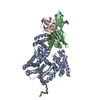
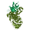
| Title | Crystal structure of Neisseria gonorrhoeae serine acetyltransferase (CysE) | ||||||
 Components Components | Serine acetyltransferase | ||||||
 Keywords Keywords |  TRANSFERASE / CysE / Serine acetyltransferase / Neisseria / Gonorrhoea TRANSFERASE / CysE / Serine acetyltransferase / Neisseria / Gonorrhoea | ||||||
| Function / homology |  Function and homology informationserine O-acetyltransferase / serine O-acetyltransferase activity / cysteine biosynthetic process from serine / cytoplasm Function and homology informationserine O-acetyltransferase / serine O-acetyltransferase activity / cysteine biosynthetic process from serine / cytoplasmSimilarity search - Function | ||||||
| Biological species |  Neisseria gonorrhoeae (bacteria) Neisseria gonorrhoeae (bacteria) | ||||||
| Method | X-RAY DIFFRACTION / SYNCHROTRON / MOLECULAR REPLACEMENT / Resolution: 2.01 Å | ||||||
 Authors Authors | Hicks, J.L. / Oldham, K.E. / Summers, E.L. / Prentice, E.J. | ||||||
| Funding support |  New Zealand, 1items New Zealand, 1items
| ||||||
 Citation Citation | Journal: Biochem.J. / Year: 2022 Title: Serine acetyltransferase from Neisseria gonorrhoeae; structural and biochemical basis of inhibition. Authors: Oldham, K.E.A. / Prentice, E.J. / Summers, E.L. / Hicks, J.L. | ||||||
| History |
|
- Structure visualization
| Structure viewer | Molecule: MolmilJmol/JSmol |
|---|
- Downloads & links
-Download
| PDBx/mmCIF format | 6wye.cif.gz | 566.2 KB | Display | PDBx/mmCIF format |
|---|---|---|---|---|
| PDB format | pdb6wye.ent.gz | 463.2 KB | Display | PDB format |
| PDBx/mmJSON format | 6wye.json.gz | Tree view | PDBx/mmJSON format | |
| Others |  Other downloads Other downloads |
-Validation report
| Arichive directory | https://data.pdbj.org/pub/pdb/validation_reports/wy/6wyeftp://data.pdbj.org/pub/pdb/validation_reports/wy/6wye | HTTPS FTP |
|---|
-Related structure data
| Related structure data |  7ra4C  3gvdS S: Starting model for refinement C: citing same article ( |
|---|---|
| Similar structure data |


-Links
 PDBj
PDBj


- Assembly
| Deposited unit | 
| ||||||||||
|---|---|---|---|---|---|---|---|---|---|---|---|
| 1 | 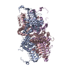
| ||||||||||
| 2 | 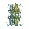
| ||||||||||
| Unit cell |
|
-Components
| #1: Protein | Mass: 31659.129 Da / Num. of mol.: 6 Source method: isolated from a genetically manipulated source Source: (gene. exp.) Neisseria gonorrhoeae (bacteria)Gene: cysE, E8M63_06755, F0T10_08440, F0T11_08395, F0T12_08425, NCTC13805_01597, WHOF_01434, WHOF_01635 Production host: Escherichia coli BL21(DE3) (bacteria) / References: UniProt: A0A1D3FX11, serine O-acetyltransferase#2: Chemical | ChemComp-NA /   Mass: 22.990 Da / Num. of mol.: 6 / Source method: obtained synthetically / Formula: Na Mass: 22.990 Da / Num. of mol.: 6 / Source method: obtained synthetically / Formula: Na#3: Chemical | ChemComp-LMR / ( Malic acid  Mass: 134.087 Da / Num. of mol.: 6 / Source method: obtained synthetically / Formula: C4H6O5 Mass: 134.087 Da / Num. of mol.: 6 / Source method: obtained synthetically / Formula: C4H6O5#4: Water | ChemComp-HOH / | Water Mass: 18.015 Da / Num. of mol.: 538 / Source method: isolated from a natural source / Formula: H2O Mass: 18.015 Da / Num. of mol.: 538 / Source method: isolated from a natural source / Formula: H2OHas ligand of interest | N | |
|---|
-Experimental details
-Experiment
| Experiment | Method: X-RAY DIFFRACTION / Number of used crystals: 1 |
|---|
- Sample preparation
| Crystal | Density Matthews: 1.92 Å3/Da / Density % sol: 36.11 % |
|---|---|
| Crystal grow | Temperature: 291 K / Method: vapor diffusion, hanging drop / pH: 6.6 / Details: 28% (v/v) Tacsimate |
-Data collection
| Diffraction | Mean temperature: 100 K / Serial crystal experiment: N |
|---|---|
| Diffraction source | Source: SYNCHROTRON / Site: Australian Synchrotron  / Beamline: MX2 / Wavelength: 0.9537 Å / Beamline: MX2 / Wavelength: 0.9537 Å |
| Detector | Type: DECTRIS EIGER X 16M / Detector: PIXEL / Date: Sep 2, 2019 |
| Radiation | Protocol: SINGLE WAVELENGTH / Monochromatic (M) / Laue (L): M / Scattering type: x-ray |
| Radiation wavelength | Wavelength: 0.9537 Å / Relative weight: 1 |
| Reflection | Resolution: 2.01→46.93 Å / Num. obs: 97704 / % possible obs: 100 % / Redundancy: 7.1 % / Biso Wilson estimate: 34.69 Å2 / CC1/2: 0.999 / Rpim(I) all: 0.023 / Net I/σ(I): 17.2 |
| Reflection shell | Resolution: 2.01→2.04 Å / Mean I/σ(I) obs: 3.5 / Num. unique obs: 4841 / CC1/2: 0.917 / % possible all: 100 |
- Processing
| Software |
| |||||||||||||||||||||||||||||||||||||||||||||||||||||||||||||||||||||||||||||||||||||||||||||||||||||||||||||||||||||||||||||||||||||||||||||||||||||||||||||||||||||||||||||||||||||||||||||||||||||||||||||||||||||||||
|---|---|---|---|---|---|---|---|---|---|---|---|---|---|---|---|---|---|---|---|---|---|---|---|---|---|---|---|---|---|---|---|---|---|---|---|---|---|---|---|---|---|---|---|---|---|---|---|---|---|---|---|---|---|---|---|---|---|---|---|---|---|---|---|---|---|---|---|---|---|---|---|---|---|---|---|---|---|---|---|---|---|---|---|---|---|---|---|---|---|---|---|---|---|---|---|---|---|---|---|---|---|---|---|---|---|---|---|---|---|---|---|---|---|---|---|---|---|---|---|---|---|---|---|---|---|---|---|---|---|---|---|---|---|---|---|---|---|---|---|---|---|---|---|---|---|---|---|---|---|---|---|---|---|---|---|---|---|---|---|---|---|---|---|---|---|---|---|---|---|---|---|---|---|---|---|---|---|---|---|---|---|---|---|---|---|---|---|---|---|---|---|---|---|---|---|---|---|---|---|---|---|---|---|---|---|---|---|---|---|---|---|---|---|---|---|---|---|---|
| Refinement | Method to determine structure: MOLECULAR REPLACEMENT Starting model: 3GVD Resolution: 2.01→43.11 Å / SU ML: 0.21 / Cross valid method: FREE R-VALUE / σ(F): 0.25 / Phase error: 25.7 / Stereochemistry target values: ML
| |||||||||||||||||||||||||||||||||||||||||||||||||||||||||||||||||||||||||||||||||||||||||||||||||||||||||||||||||||||||||||||||||||||||||||||||||||||||||||||||||||||||||||||||||||||||||||||||||||||||||||||||||||||||||
| Solvent computation | Shrinkage radii: 0.9 Å / VDW probe radii: 1.11 Å / Solvent model: FLAT BULK SOLVENT MODEL | |||||||||||||||||||||||||||||||||||||||||||||||||||||||||||||||||||||||||||||||||||||||||||||||||||||||||||||||||||||||||||||||||||||||||||||||||||||||||||||||||||||||||||||||||||||||||||||||||||||||||||||||||||||||||
| Refinement step | Cycle: LAST / Resolution: 2.01→43.11 Å
| |||||||||||||||||||||||||||||||||||||||||||||||||||||||||||||||||||||||||||||||||||||||||||||||||||||||||||||||||||||||||||||||||||||||||||||||||||||||||||||||||||||||||||||||||||||||||||||||||||||||||||||||||||||||||
| Refine LS restraints |
| |||||||||||||||||||||||||||||||||||||||||||||||||||||||||||||||||||||||||||||||||||||||||||||||||||||||||||||||||||||||||||||||||||||||||||||||||||||||||||||||||||||||||||||||||||||||||||||||||||||||||||||||||||||||||
| LS refinement shell |
| |||||||||||||||||||||||||||||||||||||||||||||||||||||||||||||||||||||||||||||||||||||||||||||||||||||||||||||||||||||||||||||||||||||||||||||||||||||||||||||||||||||||||||||||||||||||||||||||||||||||||||||||||||||||||
| Refinement TLS params. | Method: refined / Origin x: 37.491 Å / Origin y: 54.6632 Å / Origin z: 26.2118 Å
| |||||||||||||||||||||||||||||||||||||||||||||||||||||||||||||||||||||||||||||||||||||||||||||||||||||||||||||||||||||||||||||||||||||||||||||||||||||||||||||||||||||||||||||||||||||||||||||||||||||||||||||||||||||||||
| Refinement TLS group | Selection details: all |
