 Movie
Movie Controller

[English] 日本語
 Yorodumi
- PDB-6wyc: Crystal Structure of Chlamydia trachomatis Glyceraldehyde 3-phosp... -
+ Open data

- Basic information
| Entry | Database: PDB / ID: 6wyc | ||||||
|---|---|---|---|---|---|---|---|
| Title | Crystal Structure of Chlamydia trachomatis Glyceraldehyde 3-phosphate dehydrogenase | ||||||
 Components Components | Glyceraldehyde-3-phosphate dehydrogenase Glyceraldehyde 3-phosphate dehydrogenase Glyceraldehyde 3-phosphate dehydrogenase | ||||||
 Keywords Keywords | OXIDOREDUCTASE / GAPDH / NAD / Glycolysis / Chlamydia | ||||||
| Function / homology |  Function and homology informationglyceraldehyde-3-phosphate dehydrogenase (phosphorylating) / glyceraldehyde-3-phosphate dehydrogenase (NAD+) (phosphorylating) activity / glycolytic process / glucose metabolic process / NAD binding / NADP binding / cytosol Function and homology informationglyceraldehyde-3-phosphate dehydrogenase (phosphorylating) / glyceraldehyde-3-phosphate dehydrogenase (NAD+) (phosphorylating) activity / glycolytic process / glucose metabolic process / NAD binding / NADP binding / cytosolSimilarity search - Function | ||||||
| Biological species |   Chlamydia trachomatis (bacteria) Chlamydia trachomatis (bacteria) | ||||||
| Method | X-RAY DIFFRACTION / SYNCHROTRON / MOLECULAR REPLACEMENT / Resolution: 1.5 Å | ||||||
 Authors Authors | Schormann, N. / Chattopadhyay, D. | ||||||
 Citation Citation | Journal: Protein Sci. / Year: 2020 Title: Chlamydia trachomatis glyceraldehyde 3-phosphate dehydrogenase: Enzyme kinetics, high-resolution crystal structure, and plasminogen binding. Authors: Schormann, N. / Campos, J. / Motamed, R. / Hayden, K.L. / Gould, J.R. / Green, T.J. / Senkovich, O. / Banerjee, S. / Ulett, G.C. / Chattopadhyay, D. | ||||||
| History |
|
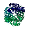
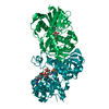
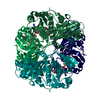
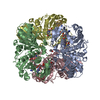
- Structure visualization
| Structure viewer | Molecule: MolmilJmol/JSmol |
|---|
- Downloads & links
-Download
| PDBx/mmCIF format | 6wyc.cif.gz | 291.8 KB | Display | PDBx/mmCIF format |
|---|---|---|---|---|
| PDB format | pdb6wyc.ent.gz | 235.4 KB | Display | PDB format |
| PDBx/mmJSON format | 6wyc.json.gz | Tree view | PDBx/mmJSON format | |
| Others |  Other downloads Other downloads |
-Validation report
| Arichive directory | https://data.pdbj.org/pub/pdb/validation_reports/wy/6wycftp://data.pdbj.org/pub/pdb/validation_reports/wy/6wyc | HTTPS FTP |
|---|
-Related structure data
| Related structure data |  6x2eC  4qx6S C: citing same article ( S: Starting model for refinement |
|---|---|
| Similar structure data |


-Links
 PDBj
PDBj
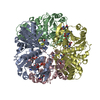
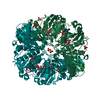
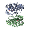
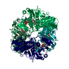
- Assembly
| Deposited unit | 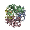
| ||||||||||||||||||||||||||||||||||||||||||||||||||||||||||||||||||||||||||||||||||||||||||||||||||||||||||||||||||||||||||||||||||||||||||||||||||||||
|---|---|---|---|---|---|---|---|---|---|---|---|---|---|---|---|---|---|---|---|---|---|---|---|---|---|---|---|---|---|---|---|---|---|---|---|---|---|---|---|---|---|---|---|---|---|---|---|---|---|---|---|---|---|---|---|---|---|---|---|---|---|---|---|---|---|---|---|---|---|---|---|---|---|---|---|---|---|---|---|---|---|---|---|---|---|---|---|---|---|---|---|---|---|---|---|---|---|---|---|---|---|---|---|---|---|---|---|---|---|---|---|---|---|---|---|---|---|---|---|---|---|---|---|---|---|---|---|---|---|---|---|---|---|---|---|---|---|---|---|---|---|---|---|---|---|---|---|---|---|---|---|
| 1 |
| ||||||||||||||||||||||||||||||||||||||||||||||||||||||||||||||||||||||||||||||||||||||||||||||||||||||||||||||||||||||||||||||||||||||||||||||||||||||
| Unit cell |
| ||||||||||||||||||||||||||||||||||||||||||||||||||||||||||||||||||||||||||||||||||||||||||||||||||||||||||||||||||||||||||||||||||||||||||||||||||||||
| Noncrystallographic symmetry (NCS) | NCS domain:
NCS domain segments: Component-ID: 0 / Beg auth comp-ID: MET / Beg label comp-ID: MET / Refine code: 0
NCS ensembles :
|
-Components
| #1: Protein | Glyceraldehyde 3-phosphate dehydrogenase / GAPDH / NAD-dependent glyceraldehyde-3-phosphate dehydrogenase Mass: 36418.605 Da / Num. of mol.: 4 Source method: isolated from a genetically manipulated source Details: The protein is modified at two cysteine positions (63 and 287). Source: (gene. exp.) Chlamydia trachomatis (strain D/UW-3/Cx) (bacteria)Strain: D/UW-3/Cx / Gene: gap, gapA, CT_505 / Plasmid: pJ411 / Production host: Escherichia coli BL21(DE3) (bacteria)References: UniProt: P0CE13, glyceraldehyde-3-phosphate dehydrogenase (phosphorylating)#2: Chemical | ChemComp-NAD / Nicotinamide adenine dinucleotide  Mass: 663.425 Da / Num. of mol.: 4 / Source method: obtained synthetically / Formula: C21H27N7O14P2 / Feature type: SUBJECT OF INVESTIGATION / Comment: NAD*YM Mass: 663.425 Da / Num. of mol.: 4 / Source method: obtained synthetically / Formula: C21H27N7O14P2 / Feature type: SUBJECT OF INVESTIGATION / Comment: NAD*YM#3: Water | ChemComp-HOH / | Water Mass: 18.015 Da / Num. of mol.: 1230 / Source method: isolated from a natural source / Formula: H2O Mass: 18.015 Da / Num. of mol.: 1230 / Source method: isolated from a natural source / Formula: H2OHas ligand of interest | Y | |
|---|
-Experimental details
-Experiment
| Experiment | Method: X-RAY DIFFRACTION / Number of used crystals: 1 |
|---|
- Sample preparation
| Crystal | Density Matthews: 1.98 Å3/Da / Density % sol: 37.8 % |
|---|---|
| Crystal grow | Temperature: 294 K / Method: vapor diffusion, hanging drop / pH: 7.5 Details: 25% PEG 2000 MME, 0.1 M Hepes, pH 7.5; prior to crystallization the protein (at 30 mg/ml) was incubated with 1 mM NAD |
-Data collection
| Diffraction | Mean temperature: 100 K / Serial crystal experiment: N |
|---|---|
| Diffraction source | Source: SYNCHROTRON / Site: APS  / Beamline: 24-ID-C / Wavelength: 0.9791 Å / Beamline: 24-ID-C / Wavelength: 0.9791 Å |
| Detector | Type: DECTRIS PILATUS 6M / Detector: PIXEL / Date: Nov 22, 2016 |
| Radiation | Protocol: SINGLE WAVELENGTH / Monochromatic (M) / Laue (L): M / Scattering type: x-ray |
| Radiation wavelength | Wavelength: 0.9791 Å / Relative weight: 1 |
| Reflection | Resolution: 1.5→66.26 Å / Num. obs: 173217 / % possible obs: 93.2 % / Redundancy: 3.3 % / Biso Wilson estimate: 12.3 Å2 / CC1/2: 0.997 / Rmerge(I) obs: 0.076 / Rpim(I) all: 0.045 / Rrim(I) all: 0.089 / Net I/σ(I): 10.2 |
| Reflection shell | Resolution: 1.5→1.53 Å / Redundancy: 3.3 % / Rmerge(I) obs: 0.626 / Num. unique obs: 8596 / CC1/2: 0.679 / Rpim(I) all: 0.388 / Rrim(I) all: 0.742 / % possible all: 93 |
- Processing
| Software |
| |||||||||||||||||||||||||||||||||||||||||||||||||||||||||||||||||
|---|---|---|---|---|---|---|---|---|---|---|---|---|---|---|---|---|---|---|---|---|---|---|---|---|---|---|---|---|---|---|---|---|---|---|---|---|---|---|---|---|---|---|---|---|---|---|---|---|---|---|---|---|---|---|---|---|---|---|---|---|---|---|---|---|---|---|
| Refinement | Method to determine structure: MOLECULAR REPLACEMENT Starting model: 4QX6 Resolution: 1.5→66.26 Å / Cor.coef. Fo:Fc: 0.965 / Cor.coef. Fo:Fc free: 0.958 / SU B: 1.618 / SU ML: 0.057 / Cross valid method: THROUGHOUT / σ(F): 0 / ESU R: 0.084 / ESU R Free: 0.079 / Stereochemistry target values: MAXIMUM LIKELIHOOD Details: HYDROGENS HAVE BEEN ADDED IN THE RIDING POSITIONS U VALUES : REFINED INDIVIDUALLY
| |||||||||||||||||||||||||||||||||||||||||||||||||||||||||||||||||
| Solvent computation | Ion probe radii: 0.8 Å / Shrinkage radii: 0.8 Å / VDW probe radii: 1.2 Å / Solvent model: MASK | |||||||||||||||||||||||||||||||||||||||||||||||||||||||||||||||||
| Displacement parameters | Biso max: 68.73 Å2 / Biso mean: 17.1 Å2 / Biso min: 5.5 Å2
| |||||||||||||||||||||||||||||||||||||||||||||||||||||||||||||||||
| Refinement step | Cycle: final / Resolution: 1.5→66.26 Å
| |||||||||||||||||||||||||||||||||||||||||||||||||||||||||||||||||
| Refine LS restraints |
| |||||||||||||||||||||||||||||||||||||||||||||||||||||||||||||||||
| Refine LS restraints NCS | Refine-ID: X-RAY DIFFRACTION / Type: interatomic distance / Weight position: 0.05
| |||||||||||||||||||||||||||||||||||||||||||||||||||||||||||||||||
| LS refinement shell | Resolution: 1.5→1.539 Å / Rfactor Rfree error: 0 / Total num. of bins used: 20
|
